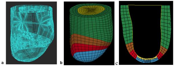
Figure 3.
Finite element model of the LV with anteroapical MI. Animal specific contours were generated from MRI (A). Solid mesh (B) was broken into four regions (green = remote, brown = BZ1 (intermediate), red = BZ2 (near MI), blue = MI) defined by wall thickness. Long-axis cross-section (C) at end-systole six-weeks post-Dor in a typical sheep (yellow = patch).

