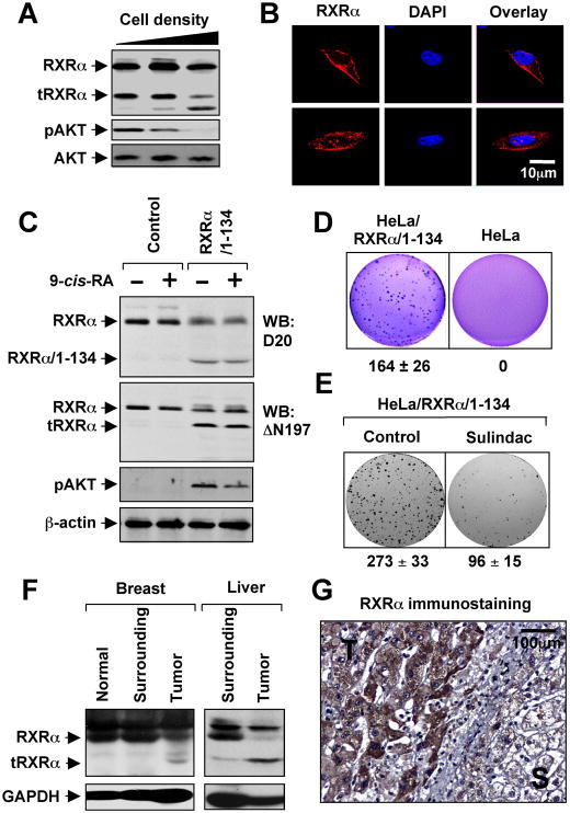
Figure 4. Role of tRXRα in AKT Activation and Anchorage-Independent Cell Growth.
(A) Cell density dependent production of tRXRα and AKT activation. MEFs seeded at different cell density were analyzed for RXRα expression using ΔN197 antibody and for AKT activation by immunoblotting.
(B) Subcellular localization of endogenous RXRα in MEFs was visualized by confocal microscopy after immunostaining using anti-RXRα (ΔN197). Cells were also stained with DAPI to visualize the nucleus. More than 60% of cells showed the images presented.
(C) Stable expression of RXRα/1–134 induces RXRα cleavage and AKT activation. HeLa or HeLa cells stably expressing RXRα/1–134 were treated with 9-cis-RA for 30 min and analyzed for AKT activation and expression of RXRα.
(D) Growth of HeLa/RXRα/1–134 and HeLa cells in soft agar.
(E) Sulindac inhibits clonogenic survival of HeLa/RXRα/1–134 cells. Cells grown in 6-well plates for 5 days were treated with Sulindac (25 μM) for 3 days.
(F) Production of tRXRα in human tumor tissues of breast (5 out of 6) or liver (4 out of 6) compared to tumor surrounding and normal tissues.
(G) Cytoplasmic localization of RXRα in liver tumor specimens immunostained by ΔN197 antibody. T, tumor tissue; S, tumor surrounding tissue.
One of three to five similar experiments is shown.
See also Figure S4.

