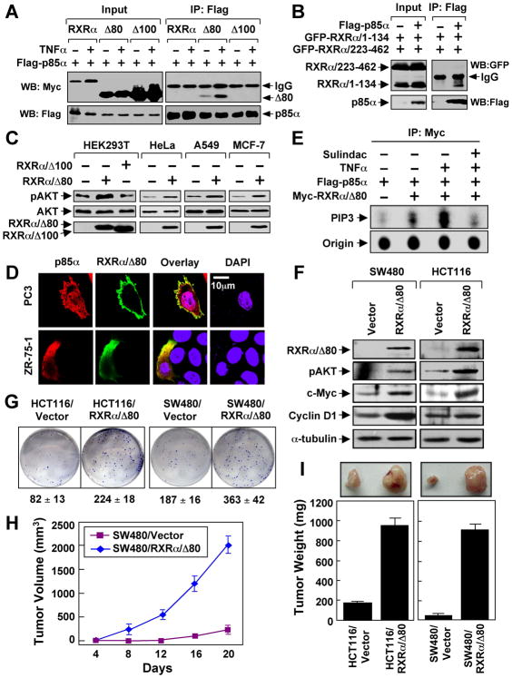
Figure 5. Role of N-terminally Truncated RXRα in PI3K/AKT Activation by TNFα and Cancer Cell Growth.
(A) HEK293T cells were transfected with Flag-p85α and RXRα, RXRα/Δ80, or RXRα/Δ100 tagged with the Myc epitope, treated with TNFα (10 ng/ml) for 30 min, and analyzed by co-immunoprecipitation using anti-Flag antibody.
(B) N-terminal A/B domain of RXRα interacts with p85α. Flag-p85α was cotransfected with GFP-RXRα/1–134 and GFP-RXRα/223–462 into HEK293T cells, and analyzed for their interaction by co-immunoprecipitation using anti-Flag antibody.
(C) RXRα/Δ80 is a potent AKT activator. AKT activation of the indicated cells transfected with RXRα/Δ80 or RXRα/Δ100 was determined by immunoblotting.
(D) Cytoplasmic co-localization of RXRα/Δ80 and p85α. Myc-RXRα/Δ80 and p85α were cotransfected into the indicated cell lines, immunostained with anti-Myc and anti-p85α antibody, and their subcellular localization revealed by confocal microscopy. RXRα/Δ80 predominantly resided in the cytoplasm of about 80% of cells. About 15% of cells showed the images presented.
(E) Activation of PI3K by RXRα/Δ80 immunoprecipitates. A549 cells transfected with Flag-p85α and Myc-RXRα/80 were treated with TNFα and/or Sulindac, immunoprecipitated with anti-Myc antibody, and subjected to in vitro PI3K assay.
(F) AKT activation by stable expression of RXRα/Δ80. Cells stably transfected with GFP-RXRα/Δ80 or control GFP vector were analyzed by immunoblotting.
(G) RXRα/Δ80 promotes clonogenic survival of cancer cells grown in 6-well plates for 8 days.
(H,I) RXRα/Δ80 promotes cancer cell growth in nude mice (n=6) for three weeks. One of three to five similar experiments is shown. Error bars represent SEM.
See also Figure S5.

