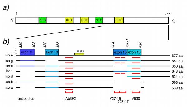
Figure 1.
Schematic representation of the FXR1 protein structure. a) Localization of the RNA-binding domains (KH and RGG; yellow) and the nuclear localization (NL) and export (NE) signals (green). b) Structure of the different FXR1P isoforms at the C-termini generated by the four small peptide inserts (in blue) deduced from the sequence of individual mRNA variants according to Kirkpatrick's et al. [18] numbering and after compilation of GenPept access No AF124386.1 to 124394.1. The boxes (exon 12, 13, 15 and 16, in blue) correspond to the peptide inserts present or absent in the different protein isoforms. The red zones indicate the regions recognized by the different antibodies.

