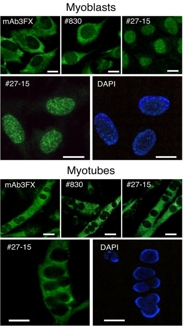
Figure 9.

Intracellular localization of the short, long and super long FXR1P isoforms by indirect immunofluorescence in myoblasts and myotubes as detected with the different antibodies to FXR1P. Confocal (upper panels) and light (lower panels) microscopy analyses after reaction with the different anti-FXR1P antibodies. Nuclei were counterstained with DAPI. Scale bars: = 5 μm.
