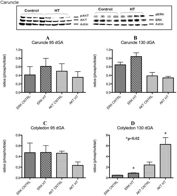
Figure 5.
Placental p-ERK and p-AKT during IUGR. A characteristic western for ERK and AKT is shown in top panel of this figure. No significant differences were observed for these proteins in the caruncle at any gestational point studies (A and B). Only a significant increase in p-ERK and p-AKT was observed in the cotyledon near term during HT treatment in the sheep (C and D).

