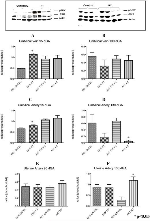
Figure 6.
Umbilical vessels and uterine artery p-ERK and p-AKT during IUGR in the sheep. Umbilical vein p-ERK was significantly increased in these vessels at mid-gestation but not near-term in the treated animals vs. controls (A and B). p-ERK was increase at mid-gestation while p-AKT was decreased in the umbilical artery of treated animals during IUGR (C and D). Uterine artery p-AKT was increase only near-term during IUGR (E and F).

