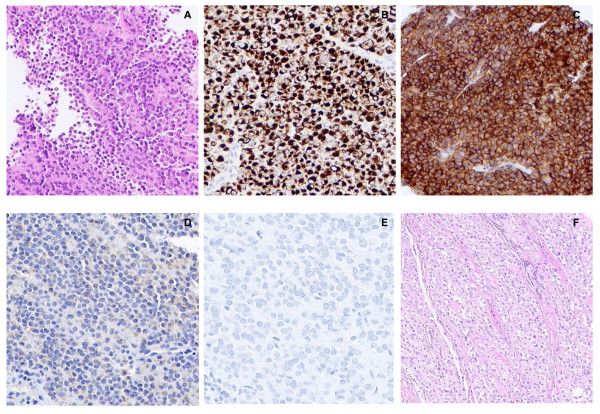
Figure 1.
Microscopic appearance and immunohistochemical features of the tumor. A) Representative area of the core needle biopsy specimen showing a homogeneous plasmacytoid appearance of the tumor cells (H&E; ×100 magnification). B) Desmin and C) CD99 strong immunoreactivity (×200 magnification). D) S100 and E) HMB-45 immunohistochemical staining (×200 magnification). F) Representative area of the resection specimen showing nests of tumor cells with clear cytoplasm divided by thin fibrous septa (×100 magnification).

