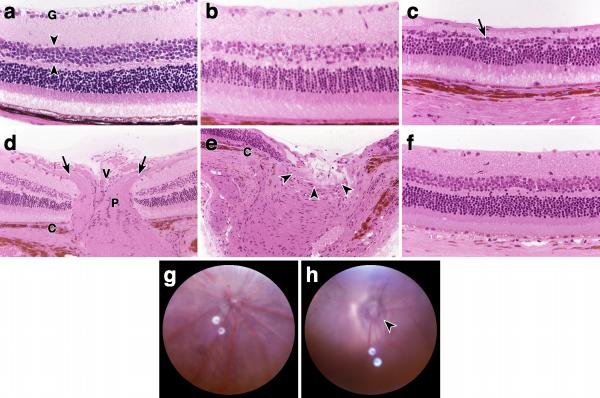
Figure 3.
Severe retinal and optic nerve damage in AKXD28 mice. The panels are arranged to display the progressive increase in severity from left to right. All images are from strain AKXD28 mice except for f, (D2) and g,h (AKXD28B6F1 X AKXD28, backcross N2). (a) Young AKXD28 retinas have normal morphology. The retinal ganglion cell layer (G) is continuous and 1-2 cells thick. The inner nuclear layer is approximately 5 to 6 cells thick (flanked by arrowheads). (b) Moderately affected AKXD28 retinas contain fewer retinal ganglion cells while the inner nuclear layer has some cell loss but remains relatively normal. (c) Severely affected AKXD28 retinas have very few retinal ganglion cells, the inner nuclear layer (arrow) is only 1-2 cells thick, and the total thickness of the retina is greatly reduced. Focal loss of photoreceptors is also present. The image represents the severe phenotype attained by all old AKXD28 eyes; some old eyes have even more cell loss with severe photoreceptor depletion and the remnants of retina are very thin. This severe atrophy does not occur in D2 mice (see f for a typical severe D2 retina). (d) Normal optic nerve head of a young mouse characterized by a thick nerve fiber layer entering the optic nerve (arrows), a central vessel (V), and well organized pial septae (P). (e) Advanced optic nerve excavation (arrowheads) with atrophy extending to a level external to the choroid (C). There is severe peripapillary atrophy with thinning of most retinal layers near the nerve. Although not prominent in this image, gliosis was frequently observed in severely damaged nerves. (f) Representative retina from a D2 mouse exhibiting advanced end stage retinal disease typical for that strain. Note that the inner nuclear layer is relatively unaffected and overall retinal thickness is maintained. (g) Normal fundus. (h) Glaucomatous fundus with an asymmetric and severely excavated optic nerve head (arrowhead). Peripapillary chorioretinal atrophy is also distinctly recognizable in this eye. These fundi are from backcross mice since all old AKXD28 mice had severe cataracts that made photography very difficult. The appearance of these backcross fundi closely resembles those of age matched AKXD28. Original magnifications 400X (a,b,c,f,) and 200X (d,e).

