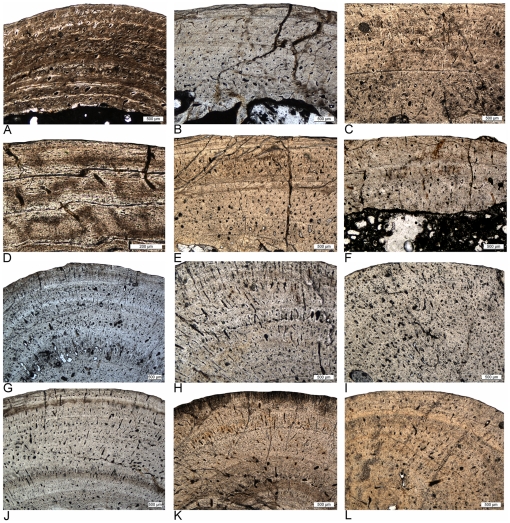
Figure 6. Different vascular density in humeri of adult Nothosaurus and Cymatosaurus (histotype A).
A–D) Samples of Nothosaurus morphotype IV humeri in normal light. Vascular density decreases from A) to C). A) Relatively high vascular density in IGWH-7, preaxial bone side. B) Moderate vascular density in IGWH-18, postaxial bone side. C) Low vascular density in IGWH-8, postaxial bone side, and D) enlargement of avascular lamellar zonal bone in IGWH-8, ventral bone side. Note in D) also the high number of flat osteocytes in the nearly avascular tissue. E–F) Samples of Nothosaurus morphotype II humeri in normal light. Vascular density decreases from E) to F). E) Relatively high vascular density in IGWH-25, preaxial bone side. F) Only moderate vascular density in IGWH-28, postaxial bone side. G–L) Samples of Cymatosaurus (histotype A) in normal light. Vascular density decreases from G) to L). G) High vascular density in IGWH-6, preaxial bone side. H) High vascular density in IGWH-1, ventral bone side. I) High vascular density in NMNHL RGM 449487, postaxial bone side. J) Moderate vascular density in IGWH-20, ventral bone side. K) Moderate to low vascular density in IGWH-19, preaxial bone side. L) Low vascular density in IGWH-29, preaxial bone side. Please note also the differences in growth marks. Whereas nothosaur humeri (A–F) have lines of arrested growth (LAGs) developed, histotype A samples (G–L) show mainly annuli.

