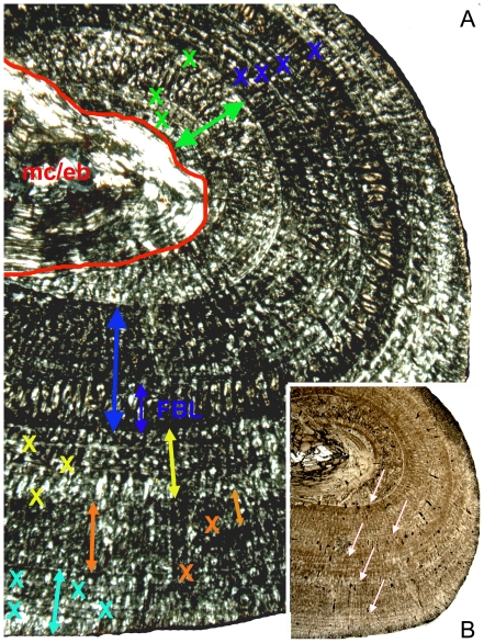
Figure 15. Growth pattern of Cymatosaurus (histotype A).
A) Postaxial bone side of humerus IGWH-19 in polarized light. The red line marks the sharp border from the by endosteally (eb) filled medullary cavity (mc) to the primary cortex. The following arrows mark the 5 main growth phases. The green arrow marks the inner phase of moderate growth. Two annuli are already developed within this inner phase of moderate growth. The large blue arrow marks the following phase of fast growth containing a thick layer of fibrolamellar bone (small blue arrow). The yellow arrow marks a phase of slow growth, consisting of well organized parallel-fibered bone tissue. The orange arrow marks a second phase of fast growth, again containing a layer of fibrolamellar bone (small orange arrow). The turquoise arrow marks another phase of slow growth. Each X marks an annulus. B) Same sample in normal light. Arrows mark the main LAGs, traceable in normal light throughout the entire cross section. Under this magnification and at this bone side are only 5 LAGs visible, whereas the same clipping under polarized light reveals altogether 16 growth marks (annuli and LAGs).

