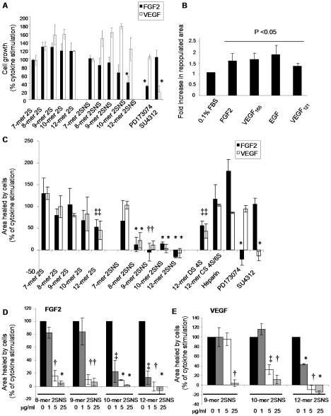
Figure 3. Oligosaccharides inhibit growth factor-induced endothelial cell proliferation and migration.
(A) Effect of oligosaccharides on endothelial cell proliferation. HUVECs were incubated with or without growth factors and oligosaccharides were added at 50 µg/ml (corresponding molar dose for each oligosaccharide is shown in Table 1), PD173074 at 0.5 µM and SU4312 at 1 µM concentration. Cell proliferation after 96 hour incubation was evaluated using SRB assay. The increase in growth factor-induced cell proliferation is expressed as 100% (control). Treatment effect is shown as a percentage of a control.Results are represented as the mean ±SD (n = 3). *, P = 0.024. (B) Cytokines stimulate endothelial cell migration. Confluent layers of serum-starved immortalized HUVECs were wounded and FGF2 (20 ng/ml), VEGF165 (20 ng/ml), EGF (20 ng/ml) and VEGF121 (20 ng/ml) were added to stimulate cell migration into the wound. The wound area at baseline and after 24 h was measured. Fold increase in the repopulated area is represented as the mean ±SD (n = 3). P<0.05. (C) Effect of oligosaccharides (all at 50 µg/ml; Table 1), heparin (50 µg/ml), PD173074 (0.2 µM) or SU4312 (0.4 µM) on the cytokine-induced repopulated wound area was tested as in B. The area that healed during 24 hours in the presence of cytokines when compared to serum-starved cells is expressed as 100%. The effect of oligosaccharides, heparin and receptor tyrosine kinase inhibitors is expressed as a percentage of repopulated area induced by a cytokine alone. The mean ± SD (n = 3) is shown. *, P<0.0001; †, P<0.001; ‡, P<0.005. (D–E) Inhibition of FGF2- or VEGF165-induced wound closure at increasing concentrations of oligosaccharides that inhibit cell migration by more than 80% at a maximal 50 µg/ml concentration. The mean ± SD (n = 2) is shown. *, P<0.001; †, P<0.005; ‡, P<0.05.

