Abstract
Background
Many developmental genes are still active in specific tissues after development is completed. This is the case for the homeobox gene Otx2, an essential actor of forebrain and head development. In adult mouse, Otx2 is strongly expressed in the retina. Mutations of this gene in humans have been linked to severe ocular malformation and retinal diseases. It is, therefore, important to explore its post-developmental functions. In the mature retina, Otx2 is expressed in three cell types: bipolar and photoreceptor cells that belong to the neural retina and retinal pigment epithelium (RPE), a neighbour structure that forms a tightly interdependent functional unit together with photoreceptor cells.
Methodology/Principal Findings
Conditional self-knockout was used to address the late functions of Otx2 gene in adult mice. This strategy is based on the combination of a knock-in CreERT2 allele and a floxed allele at the Otx2 locus. Time-controlled injection of tamoxifen activates the recombinase only in Otx2 expressing cells, resulting in selective ablation of the gene in its entire domain of expression. In the adult retina, loss of Otx2 protein causes slow degeneration of photoreceptor cells. By contrast, dramatic changes of RPE activity rapidly occur, which may represent a primary cause of photoreceptor disease.
Conclusions
Our novel mouse model uncovers new Otx2 functions in adult retina. We show that this transcription factor is necessary for long-term maintenance of photoreceptors, likely through the control of specific activities of the RPE.
Introduction
The molecular basis of some forms of retinitis pigmentosa [1] and of late onset retinal degeneration, in particular age-related macular degeneration (AMD) remains obscure. Studying these diseases requires animal models designed to explore gene function in adults. Unfortunately, such tools are still largely unavailable [2]. Knockout models often result in deficiencies that occur during development rather than in mature retina, precluding assessment of gene function in adults.
One transcription factor that is expressed in retinal cell types (photoreceptors and retinal pigment epithelium), where abnormal function can lead to various diseases, is the homeoprotein Otx2. Otx2 is well studied for its role in embryonic head formation [3], and differentiation of various types of neurons [4], [5]. It has also been proposed to modulate the plasticity of the visual cortex through a paracrine mechanism [6]. Recent studies have demonstrated its requirement at many steps of eye development. Otx2 first acts at mid-gestation allowing the differentiation of the RPE territory [7], [8]. It is then necessary for neural retina development, maturation and photoreceptor fate determination [9], and for bipolar cell terminal differentiation after birth [10], [11]. These studies have proven very informative for the role of Otx2 in development of the retina. Still, little is known about its function in the adult, although it is strongly expressed in the adult retina [10], [12], [13].
Some patients that suffer from microphthalmia and anophthalmia have been reported to carry OTX2 mutations [14]. It was also noted that two eye diseases that are likely unrelated to development, retinitis pigmentosa (RP) and Leber congenital amaurosis (LCA) sometimes developed in these patients. It has also recently been reported that Otx2, and Crx, are highly expressed in retinoblastoma tumor cells in patients [13]. The Otx2 gene uses three different promoters and has many enhancers that act at different times and cellular types spread over 300 kb [15], [16], [17]. Therefore it is quite possible that changes in noncoding sequences regulating Otx2 transcription might lead to retinal disease. To explore the functions of Otx2 in the adult eye, we took advantage of the novel genetic approach coined self-knockout that we previously used to study Otx2 early functions [18] and which allows time-controlled and very efficient knockout of Otx2 only in cells that express it.
Results
Evaluation of Otx2 knockout in the adult retina
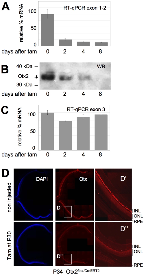
Sustained expression in RPE, photoreceptors and bipolar cells (BC) (Fig. S1) suggested that Otx2 gene could exert functions in the mature retina. In order to address this issue, we used Otx2flox/CreERT2 adult mice (Fig. S2). In this model, tamoxifen inducible CreERT2 recombinase is expressed from one Otx2 allele whereas the other allele carries two loxP sites flanking the homeodomain coding exon 2. The floxed allele expresses normal levels of Otx2 protein. As the CreERT2 gene is inserted at the Otx2 locus, tamoxifen treatment induces CreERT2-mediated deletion of the functional allele only in cells that express Otx2, a strategy we termed self-knockout [18]. We triggered Otx2 self-knockout in P30 Otx2flox/CreERT2 mice. Gene ablation in the whole retina was assessed by measurement of mRNA and protein abundance during 8 days following tamoxifen treatment. Quantitative RT-PCR (RT-qPCR) analysis of full length Otx2 transcript revealed a reduction of 90 to 95% of its abundance after 2 days. This level remained stable at later times (Fig. 1A). Western blot analysis also showed a decrease of Otx2 protein levels (Fig. 1B), which became undetectable 8 days after tamoxifen injection. Consistently, in histological sections, Otx2 expression in the RPE, photoreceptor layer and inner nuclear layer appeared dramatically reduced throughout the retina 4 days following tamoxifen treatment (Fig. 1D). However, transcription of the Otx2 locus after self-knockout was not affected: quantification of deleted and full length mRNAs by RT-qPCR using exon 3 primers (Fig. 1C) and Cre mRNA by in situ hybridization (Fig. S3, right panel) revealed a steady-state level of mRNA, ruling out Otx2 autoregulation in the mature retina. Together, these results show that triggering self-knockout in adult retina leads to an almost complete Otx2 protein loss within 8 days.
Figure 1. Efficient loss of Otx2 gene products after self-knockout in adult retina.
A–C. Time course analysis of Otx2 gene products following tamoxifen injection at P30 in Otx2flox/CreERT2 mice. Relative levels of full-length (A) and total (normal + deleted) (C) Otx2 mRNA measured by RT-qPCR are shown. Corresponding levels of Otx2 protein (B) is shown in western blots. Position of size markers and Otx2 protein are indicated. Error bars in A and C are standard deviation. D. Staining of nuclei (DAPI) and Otx proteins on identical retina sections from control and mutant mice of the same genotype 4 days after tamoxifen injection. Shown are stitching of overlapping fields reconstituting a whole section. Right panel: magnification of boxed D′ and D″ areas. RPE: retinal pigment epithelium; ONL: outer nuclear layer; INL: inner nuclear layer.
Alterations of photoreceptors
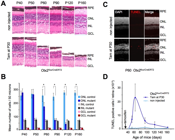
The consequences of Otx2 gene ablation in adult retina were examined in a series of mice, from P40 to P160, corresponding to 10–130 days after treatment (Fig. 2A). In Otx2flox/CreERT2 retina treated with tamoxifen at P30, we observed first a shortening of the photoreceptor outer segments, which were detached from the RPE from about P50. With time, the outer and inner segments progressively disappeared until by P120 they were altogether entirely absent. The whole retina was affected. In parallel we observed a progressive thinning of the outer nuclear layer, which started from around P60, until, by P160 no or extremely few photoreceptors were left. We also noted that with time from the injection of tamoxifen, the retina was increasingly detached from the RPE – although this likely was exacerbated by the histological procedure. Control Otx2flox/CreERT2 animals exhibited normal retinal histology. Retinas of tamoxifen-injected Otx2flox/flox and Otx2+/CreERT2 mice appeared similar to control retinas (Fig. S4) ruling out any potential nonspecific toxicity of tamoxifen treatment. Cell counting revealed progressive disappearance of photoreceptors and no change of cell numbers in both inner and ganglion cell layers (Fig. 2B). The kinetics of photoreceptor loss was identical in central and peripheral retina as well as in dorsal and ventral retina.
Figure 2. Loss of Otx2 leads to photoreceptor cell degeneration.
A. Histology of control (non injected) and mutant (Tamoxifen administrated at P30) series of Otx2flox/CreERT2 retinas of the indicated ages. Sections are stained with Eosin and Haematoxylin. RPE: retinal pigment epithelium; ONL: outer nuclear layer; INL: inner nuclear layer; GCL: ganglion cell layer. B. Cell counts in the three layers retinal sections of control or tamoxifen treated (mutant) Otx2flox/CreERT2 mice at indicated ages. Normalized fields of the same eye area were used. Mean cell number and standard deviation are indicated for each condition (*P<0.001). C. Detection of apoptotic cells in control or tamoxifen treated Otx2flox/CreERT2 retinas 30 days after treatment. Left, middle and right panels show respectively DAPI staining, TUNEL labelled cells and superimposition of both images. D. Kinetics of apoptosis following Otx2 gene ablation. TUNEL labelled cells were counted on normalized sections of three independent mice for each stage, each corresponding to 0.063 mm2 of retinal area. Error bars are standard deviation.
To investigate more closely the reason and time-course of photoreceptor disappearance we did TUNEL staining on a series of retinas from the same ages. Positive TUNEL cells could only be found in the photoreceptor layer (Fig. 2C). We studied the kinetics of apoptosis by quantifying TUNEL staining at various timepoints following tamoxifen injection. Retinas from treated mice had few apoptotic photoreceptors at P40, 10 days following tamoxifen injection. However the numbers of dying photoreceptors increased very much by P50, reaching a maximum around P60 (30 days after injection), and then slowly decreasing until P160, by which time virtually no photoreceptors remained (Fig. 2D). This kinetics was consistent with the observed decrease of thickness of the outer nuclear layer. In control mice there were very few apoptotic cells and the thickness of the outer nuclear layer remained unchanged throughout this period. Thus, Otx2 depletion in adult retina leads to selective and slow photoreceptor cell death by apoptosis.
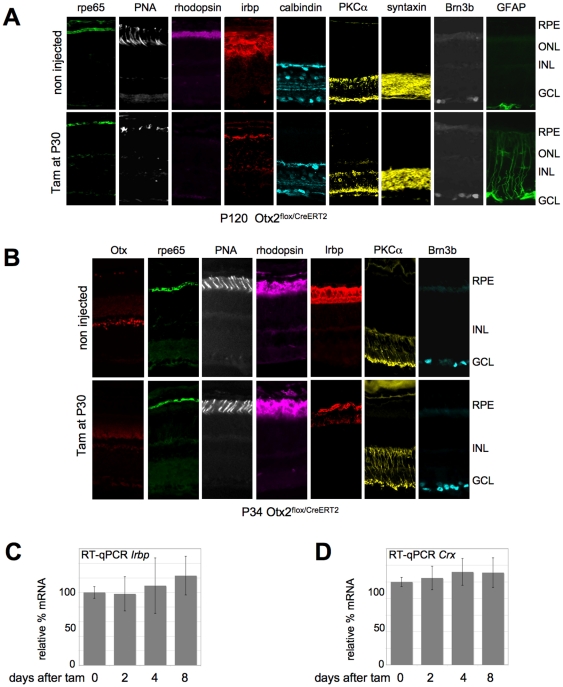
Consistent with photoreceptor alteration, PNA labelling of cone outer segments was reduced in mutants, which also showed absence of rhodopsin at P120 (Fig. 3A). IRBP, a glycoprotein of the inter-photoreceptor matrix synthesized by photoreceptors, was less abundant in mutants. We also investigated expression of cell-type specific markers in P120 mutant retinas. Syntaxin labelling indicated no expansion of the amacrine population. Expression of markers of RPE (rpe65), horizontal (calbindin), bipolar (PKC-alpha, calbindin) and ganglion cells (Brn3b) also did not appreciably change in mutant retina. However, GFAP labelling of Müller cells was enhanced in mutant retinas, a hallmark of glial reaction typical of degenerating neural tissue [19].
Figure 3. Consequences of Otx2 loss in photoreceptor cells.
A. Expression of cell type specific markers in P120 control (upper panels) and knockout (lower panels) retinas, following tamoxifen treatment at P30. B. Expression of cell type specific markers in P34 control (upper panels) and self-knockout (lower panels) retinas, following tamoxifen treatment at P30. C. Relative levels of Irbp mRNA in Otx2flox/CreERT2 retina following tamoxifen treatment. D. Relative levels of Crx mRNA in Otx2flox/CreERT2 retina following tamoxifen treatment. Error bars are standard deviation.
To find out if expression of some of the above markers was directly linked to that of Otx2, we analyzed their expression soon after tamoxifen injection. Four days after treatment, although Otx2 protein was strongly reduced, we observed no change in expression of most tested markers including rhodopsin (Fig 3B), with one exception, IRBP whose staining appeared decreased. This prompted us to analyze Irbp gene expression. Although in vitro experiments have suggested it to be regulated by Otx2 [20], we found no change in Irbp mRNA level by RT-qPCR (Fig. 3C), suggesting that its decrease in sections was either a secondary consequence of other alterations, or that it was not due to transcriptional regulation. Crx, a major determinant of photoreceptor gene expression, appeared another interesting candidate [21]. Otx2 protein is required during embryogenesis to activate Crx in photoreceptor precursors [9] and this might also be needed during postnatal development [10]. However, we failed to observe any change in Crx mRNA level either by in situ hybridization 30 days after tamoxifen administration (Fig. S5) or by RT-qPCR immediately following tamoxifen treatment (Fig. 3D). A panel of photoreceptor specific genes – Gnat2, Rhodopsin, Grk1, Opsin1, Pde6b - was also tested by RT-qPCR. None displayed any change over 8 days following knockout (6) showing no dramatic functional alteration of the photoreceptor itself upon Otx2 gene ablation.
To summarize, these observations suggest that knockout of Otx2 gene induces a specific loss of photoreceptors, which starts with a lag of about 2 weeks following the knockout and then continues during about 4 months, until no photoreceptors remain. Our observations also show a progressive shortening and degeneration of the outer segments of photoreceptors during this period. Other retinal cell types are not affected to any comparable degree.
Functional alteration of retinal pigment epithelium
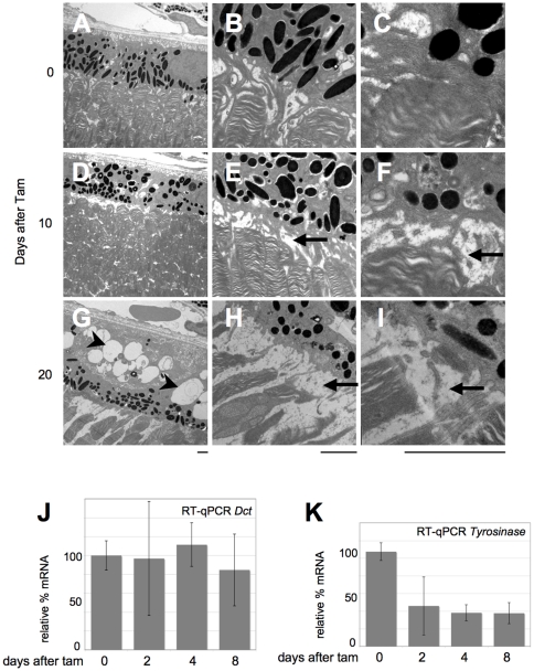
The retinal pigment epithelium expresses Otx2 at a high level, and is necessary for function and maintenance of the photoreceptors. To investigate morphological consequences of Otx2 knockout, both on RPE cells and on photoreceptor outer segments, we performed electron microscopic analysis of control and mutant retina 10 and 20 days post tamoxifen treatment. We found rapid, progressive and severe alterations of RPE cells. Ten days after knockout, RPE cells already appeared thinner with smaller and less numerous melanosomes (Fig. 4D–F). In parallel, we found disruption of RPE contacts with photoreceptor outer segments, which resulted in widening of the inter-photoreceptor matrix space (Fig. 4E, F). This worsened in retinas 20 days post treatment, with an almost complete loss of RPE contacts with disc-containing photoreceptor outer segments (Fig. 4G–I). In addition, the melanosome and melanin content of RPE cells appeared reduced, suggesting a block in biogenesis or maintenance of this cell compartment. Low-power views also revealed two important RPE modifications: i) relocalization of the remaining melanosomes. Whereas evenly dispersed in control RPE cells (Fig. 4A), these accumulated at the apical cell side 20 days post treatment (Fig. 4G). ii) extensive vacuolization of RPE cells (Fig. 4D, G). This became very important at P50 (Fig. 4G) suggesting strong metabolic reaction such as autophagy [22].
Figure 4. Rapid induction of RPE disease by Otx2 self knockout.
A–I. Transmission electron microscopy images of control (A–C) and self-knockout Otx2flox/CreERT2 retina 10 days (D–F) and 20 days (G–I) after tamoxifen injection. Low (A,D,G), medium (B,E,H) and high (C,F,I) power views are presented, focusing on the area of interaction between RPE and photoreceptor disk-containing outer segments. Scale bars: 2 µm. J–K. Relative levels of Dct (J) and tyrosinase (K) mRNA in Otx2flox/CreERT2 retina following tamoxifen treatment. Error bars are standard deviation.
As melanin amount decreased in Otx2 mutant retinas, we investigated expression of genes involved in its synthesis. Tyrosinase and DOPAchrome tautomerase (DCT) control two critical steps of melanogenesis, and Otx2 can transactivate both genes' promoter in vitro [7], [23]. We therefore tested the status of Dct and tyrosinase mRNA in mutants by RT-qPCR. Whereas Dct expression appeared unchanged (Fig. 4J), tyrosinase mRNA accumulation decreased (Fig. 4K), with kinetics that strictly paralleled the one of Otx2 mRNA shown in Fig. 1A.
We conclude from these data that Otx2 knockout in adult retina results in disruption of photoreceptor-RPE cell adhesion and in impaired melanogenesis in RPE cells. These defects as well as downregulation of tyrosinase start soon after knockout.
Discussion
Our observations demonstrate that Otx2 knockout induces profound alterations of RPE and photoreceptor cells, in adults. In our model, the most conspicuous retinal phenotype was the degeneration of photoreceptor cells. This process was very progressive, lasting for about 4 months following knockout, but ended in complete loss of photoreceptors. Otx2 knockout also resulted in profound alterations of RPE such as reduction of melanosome size and number, which were evident at a much earlier time than most photoreceptor alterations.
Several studies have previously shown a role of Otx2 in early or late development of RPE, retina, and in the differentiation of some retinal cell types [7], [8], [9], [10], [11], [14], [24]. Our study demonstrates an important role of this gene in adult retina. This is due to our use of a time-controlled Cre line, which allows Otx2 invalidation, with very high efficiency, in all cells that express Otx2. To distinguish unambiguously defects that are cell-autonomous from those that result from cell-cell interactions, it will be necessary to trigger Otx2 knockout only in restricted cell types in the adult. This will require other inducible Cre lines –e.g. for photoreceptor, bipolar, or RPE cells– or alternative experimental approaches. Several retinal cell-specific Cre lines have being made available, but most of them are not inducible or use promoters that are not efficiently expressed in adults. For instance, the RPE-specific Mct3-CreERT2 transgenic mouse described recently causes recombination only in 5% of RPE cells in adults [25]. On the other hand our technology provides for a quasi complete knockout of Otx2, which for some purposes (among which include a considerably greater ease in analyzing changes in gene expression induced by loss of Otx2 function) is advantageous.
From our study it seems clear that Otx2 plays a direct role in maintaining the integrity of RPE cells and in one of their major functions: melanogenesis. This is because knockout of Otx2 results in a quick alteration of these cells, and reduction of their melanosome content. Such a reduction exposes mutant RPE cells to oxidative damages. As these induce an increase in intracellular vacuoles [26], they could explain the vacuolization we observe in RPE cells 20 days after Otx2 gene invalidation. In addition we observe a synchronous decrease of tyrosinase and Otx2 mRNA after tamoxifen treatment. In avian retina cells, Otx2 induces a pigmented phenotype and in vitro, the protein binds to a bicoid site present in the promoter of the tyrosinase gene [7]. This adds evidence in favour of tyrosinase as an Otx2 direct target gene in vivo, although direct proof of this will be required. Given the importance of RPE cells in nurturing and maintenance of photoreceptors, impairment of RPE functions could largely explain the slow photoreceptor degeneration that we observed. Melanosomes protect the retina from photo-oxidation that can be intense in RPE which lies against the oxygen rich choriocapillaris and receives focused light [27]. Another fast change in RPE cells is the widening of the subretinal space between RPE apical microvilli and photoreceptor outer segments. This might impair at least three RPE functions: trans-epithelial transport, visual cycle and phagocytosis, a condition that can lead to retinal degeneration [28]. It could involve adhesion molecules and extra-cellular matrix components. For instance, Ezrin mutants present reductions in the apical microvilli and basal infoldings in RPE cells [29]. Disruption of RPE-photoreceptor cell adhesion could also affect the homeostasis of the inter-photoreceptor matrix, which could explain the observed reduction of IRBP protein despite normal transcription of the Irbp gene.
Alternatively, or additionally, a slow cell-autonomous degeneration could be induced after Otx2 knockout in photoreceptor cells, independent of RPE abnormalities. Although on the whole we detected severe disorders in photoreceptors following an appreciable lag compared to changes within RPE, some defects were noted in photoreceptors soon after Otx2 knockout: 10 days after treatment there already appeared alterations in the outer segments of photoreceptors at their juncture with RPE (only visible with electron microscopy), and the loss of IRBP, likely reflecting a loosening of the matrix around photoreceptors, was already visible 4 days after tamoxifen treatment. Both changes however might be a consequence of defects autonomous to RPE cells, rather than to photoreceptors. It is remarkable that TUNEL staining detected apoptotic photoreceptor cells only 20 days following tamoxifen treatment. It is also possible, that our procedure resulted in incomplete knockout of Otx2 in photoreceptors. As Otx2 seems to be expressed at a lower level there, the Cre recombinase might also be expressed at a low level which would decrease the fraction of cells in which it will act. We think this possibility unlikely because we observed a very high rate of recombination and a very strong reduction in Otx2 expression in all instances where we have used this strategy [18]. Moreover, we have noted before that in photoreceptor cells, Otx2 is concentrated in the periphery of the nucleus [12] – in other words, each cell could express a high level of Otx2 in a relatively small subcellular compartment.
It therefore seems plausible that the severe alterations in RPE cells, which express a high level of Otx2, caused the process of photoreceptor degeneration. At the same time we cannot exclude that disruption of Otx2 function might result in a delayed and staged schedule of apoptosis of these cells. This interesting possibility remains to be investigated - we note that in knockout mice for another homeoprotein, Engrailed, there occurs also a progressive loss of dopaminergic neurons [30]. The use of cell-specific inducible Cre mice will in any case allow us to address these issues in the future.
Although the main focus of this study was not to investigate which genes are downstream of Otx2, our results suggest that embryonic and adult Otx2 protein functions differ greatly. For instance although Crx expression is controlled by Otx2 in precursors of photoreceptor cells [31] we find that this is no longer the case in adult retina. This is of special interest as many retinal degenerative diseases are not developmental pathologies. They occur in adults and may be the consequence of somatic mutations or gene dosage in patients carrying heterozygous mutations [14]. It will be important to discover what are the targets of Otx2 in the mature retina and to look for de novo OTX2 mutations in patients suffering specifically from late onset retinal diseases. The subset of OTX2 mutations that should be expected would likely involve promoter or cis-regulatory change. Indeed, mutations affecting OTX2 protein coding sequence have early and severe effects on brain and eye development [14], [24]. Finding mutations outside the coding sequence may be difficult as regulatory elements of this small gene are scattered over large distances [16], [17]. However, we previously showed that Otx2 gene is expressed from three different stage- and tissue-specific promoters, the second of them accounting for most of retinal transcripts in adults [15], [32]. Mutations affecting this promoter could therefore specifically reduce the amount of Otx2 protein in the retina without affecting brain and eye development.
Methods
Mouse breeding and tamoxifen administration
Generation of mouse lines (129Sv background) and genotyping were done as previously described [12], [18]. Mice were housed with a 12 h light/dark cycle. One intraperitoneal injection of tamoxifen (Sigma-Aldrich, St Louis, MO, USA) (10 mg/ml in sunflower oil) was done at 1 mg per 20 g body weight at 3:00 pm. All experiments were conducted under guidelines approved by local and state ethical committees. TL received the authorization to experiment #69101850 from the Veterinarian Service of the Préfecture du Rhône, France, and protocols used in this study were approved by the Comité Régional d'Ethique Rhône-Alpes (decision #0175).
Immunocytochemistry
Eyes were fixed in 4% paraformaldehyde in phosphate-buffered saline (PBS) for 3 hours at 4°C, rinsed twice (30 min) in PBS, protected in PBS-sucrose (10–30%) and frozen in Tissue-Tek OCT at −80°C. Sections (8 µm) were mounted onto SuperFrost + slides (Fisher Scientific, Atlanta, GA), blocked 1 hour in PBT (PBS with 0.2% Gelatin and 0.1% Triton X-100) containing 5% donkey serum and incubated overnight at 4°C with the primary antibodies diluted in PBT with 2% donkey serum. After rinsing, slides were incubated 1 hour at 20°C with the secondary antibodies. Specimens were washed twice 15 min in PBS, coverslipped with Vectashield, observed under fluorescent Axioplan microscope (Carl Zeiss, Germany) and images analysed using Metamorph (Molecular Devices, Sunnyvale, CA, USA).
Antibodies and working dilutions. Primary antibodies: Monoclonal anti-RPE65 (1/500) (Abcam, Cambridge, UK), anti-syntaxin (1/1,000) (Sigma-Aldrich), anti-PKCalpha (1/1,000) (Santa Cruz Biotechnology; Santa Cruz CA, USA), anti-calbindin (1/500) (Sigma-Aldrich); Rabbit anti-rhodopsin (1/1,000) (Sigma-Aldrich); Goat anti-Brn-3 (1/100) and Goat anti-GFAP (1/200) (Santa Cruz Biotechnology); Rabbit anti-IRBP (1/200) (kind gift from B. Wiggert's lab); Goat anti-Otx2 (1/250) (R&D systems, Minneapolis, MN, USA) and Rat anti-Otx (1/200) (kind gift from M. Wassef).
Secondary antibodies (1/1,000): Donkey anti-Mouse Alexa Fluor 488, anti-Mouse Alexa Fluor 647, anti-Goat Alexa Fluor 647, anti-Rat Alexa Fluor 488 and anti-Rabbit Alexa Fluor 555 (Invitrogen, Carlsbad CA, USA). Cone staining used FITC-labeled peanut agglutinin (PNA, generous gift of O. Dkhissi-Benyahya).
Histological studies and cell counting
Eyes were processed as above. Sections were stained with haematoxylin and eosin according to standard protocol. Cell numbers were determined by counting the nuclei in a 50 µm wide region of retinal section located at equal distance from the ora serrata and the optic disk. For each point, three eyes were dissected. For each, three different regions were counted. Average cell numbers and standard deviation were calculated using Statlab (SPSS Inc, Chicago, Illinois, USA). TUNEL assay was performed with the In Situ Cell Death Detection Kit, TMR red (Roche, Basel, Switzerland), according to the manufacturer's instructions.
RNA Isolation and RT-qPCR
Retina were dissected at 3:00 pm in PBS. Total RNA was prepared with TRIzol (Invitrogen). First-strand cDNA was synthesized using 1 µg of total mRNA, MLV reverse transcriptase (Promega, Madison WI, USA) and random hexamers. For real time PCR, 1/100 of cDNA was used per reaction using PowerSYBR green PCR mix and StepOne Plus apparatus and software (Applied Biosystems, Foster City, CA, USA). Comparable efficiency of each PCR reaction was first demonstrated using serial dilutions of control cDNA. Gene to TBP ratios were determined from three independent assays by the 2−DCt method [33]. PCR conditions and primers used: PCR cycles: 15 s at 95°C, 30 s at 60°C, 30 s at 72°C. Primers forward and reverse (amplicon size):
Otx2 exon 1–2: 5′-ccaaatctacccaccaagga, 5′-agagcttccagaacgtcgag (276 bp)
Otx2 exon 3: 5′-GCTGGCTCAACTTCCTACT, 5′-TCCAAGCAGTCAGCATTGAAG (260 bp)
Tyrosinase: 5′-ATTGATTTTGCCCATGAAGCA, 5′-TTCCATCGCATAAAACCTGAT (283 bp)
Crx: 5′-TGTATGCACGTGAGGAGGT, 5′-GCTGGACTCCAAATGGACA (301 bp)
Irbp: 5′-GGACCCACACTCAGCTCA, 5′-AGACAACTACATTAGGTGTCA (285 bp)
Dct: 5′-GCTACAATTACGCCGTTGAT, 5′-TTCCACCTGTCTCAAGATGA (268 bp)
TBP: 5′-TCTGGAAAAGTTGTATTAACAG, 5′-GCTGCAGGGTGATTTCAGT (284 bp)
Gnat2: 5′- TCTGTTTTCCGGAGTATGAC, 5′- TGCATGAAGCCTCAGATTCT (270 bp)
Rho: 5′- TGTGGCCTTCTACATCTTCA, 5′- ACAGGAGACTCCTACTTTCA (278 bp)
Grk1: 5′- AGCTGGAGGCAGCTCGAA, 5′- ACATTCCCGACTTGGCTGT (307 bp)
Opn1: 5′- CTACTGCTTCATGAATAAGCA, 5′- GAAGGTGACTCACCAGAGT (266 bp)
Pde6b: 5′- CGATTTCACGAAGAGATCCT, 5′- TAGGCAGAGTCCGTATGCA (271 bp)
Western blot
Retinas were dissected in cold PBS and nuclear protein extracts [34] were prepared as described previously [35]. Proteins (30 µg) were fractionated on 10% SDS-PAGE and transferred onto nitrocellulose (Optitrans, Schleicher & Schuell, Dassel, Germany) and incubated with rat anti-Otx antibody (1/1,000). Revelation was done using HRP-coupled goat anti-rat antibody (1/10,000) (Jackson ImmunoResearch, West Grove, PA, USA).
Transmission electron microscopy (TEM)
Mice were perfused transcardially under deep anaesthesia with PBS, then fixative (1% paraformaldehyde, 3% glutaraldehyde in phosphate buffer 0.1 M, pH 7,4). Eyes were fixed 2 h at 4°C. Corneas were removed and eyes fixed 2 h. Lens were removed and eyecups fixed 2 h. Ten 1×1 mm retinal strips were processed separately. Samples were osmicated, dehydrated with ethanol series. Each sample was embedded in Epon mixture and 0.5 µm thin sections were stained with toluidine blue for light microscopy followed by ultra thin section preparation for TEM (Philips CM120, Eindhoven, NL).
Supporting Information
Otx2 protein in adult retina. Expression of Otx2-GFP protein in adult (P60) retina of Otx2+/Otx2−GFP mouse. Direct protein fluorescence (green) of a vertical section is shown. RPE: retinal pigment epithelium; ONL: outer nuclear layer; INL: inner nuclear layer; GCL: ganglion cell layer.
(0.64 MB TIF)
The self knockout strategy. The structure of alleles used for self knockout is presented at the top with Otx2 exons as boxes (gray: non coding, light blue: coding regions, dark blue: homeobox), knock-in CreERT2 (yellow box) and neo (blue box) genes and loxP sites (red triangles). Black arrows are transcription start sites. PCR primer pairs are blue and purple arrows. The structure resulting from tamoxifen-induced CreERT2 activity is shown at the bottom.
(0.26 MB TIF)
Otx2 driven CreERT2 expression is stable in the absence of Otx2. In situ hybridization was performed 30 days post injection on retina of control or tamoxifen injected Otx2flox/CreERT2 mice using the whole CreERT2 coding sequence as a probe. RPE: retinal pigment epithelium; ONL: outer nuclear layer; INL: inner nuclear layer; GCL: ganglion cell layer.
(0.71 MB TIF)
Absence of tamoxifen toxicity. Histology of P160 control and treated (Tamoxifen administrated at P30) retinas of the indicated genotypes. Sections are stained with Eosin and Haematoxylin. RPE: retinal pigment epithelium; ONL: outer nuclear layer; INL: inner nuclear layer; GCL: ganglion cell layer.
(0.80 MB TIF)
Crx expression is independent of Otx2 in adult retina. In situ hybridization was performed on P60 sections of control and mutant retina after tamoxifen treatment at P30 with full-length Crx cDNA (a kind gift of C. Cepko) as a probe. RPE: retinal pigment epithelium; ONL: outer nuclear layer; INL: inner nuclear layer; GCL: ganglion cell layer.
(0.44 MB TIF)
Stable expression of several photoreceptor markers following Otx2 self-knockout. RT-qPCR quantization of mRNA abundance was performed with the indicated photoreceptor specific genes. Shown is the mean of three independent experiments. Error bars are standard deviation.
(0.36 MB TIF)
Footnotes
Competing Interests: The authors have declared that no competing interests exist.
Funding: This work was supported by a Centre National de la Recherche Scientifique grant to FF and TL and a Retina France grant to TL. NF was supported by a fellowship from Retina France. The funders had no role in study design, data collection and analysis, decision to publish, or preparation of the manuscript.
References
- 1.Bok D. Contributions of genetics to our understanding of inherited monogenic retinal diseases and age-related macular degeneration. Arch Ophthalmol. 2007;125:160–164. doi: 10.1001/archopht.125.2.160. [DOI] [PubMed] [Google Scholar]
- 2.Lavado A, Montoliu L. New animal models to study the role of tyrosinase in normal retinal development. Front Biosci. 2006;11:743–752. doi: 10.2741/1832. [DOI] [PubMed] [Google Scholar]
- 3.Acampora D, Gulisano M, Broccoli V, Simeone A. Otx genes in brain morphogenesis. Prog Neurobiol. 2001;64:69–95. doi: 10.1016/s0301-0082(00)00042-3. [DOI] [PubMed] [Google Scholar]
- 4.Puelles E, Acampora D, Lacroix E, Signore M, Annino A, et al. Otx dose-dependent integrated control of antero-posterior and dorso-ventral patterning of midbrain. Nat Neurosci. 2003;6:453–460. doi: 10.1038/nn1037. [DOI] [PubMed] [Google Scholar]
- 5.Puelles E, Annino A, Tuorto F, Usiello A, Acampora D, et al. Otx2 regulates the extent, identity and fate of neuronal progenitor domains in the ventral midbrain. Development. 2004;131:2037–2048. doi: 10.1242/dev.01107. [DOI] [PubMed] [Google Scholar]
- 6.Sugiyama S, Di Nardo AA, Aizawa S, Matsuo I, Volovitch M, et al. Experience-Dependent Transfer of Otx2 Homeoprotein into the Visual Cortex Activates Postnatal Plasticity. Cell. 2008;134:508–520. doi: 10.1016/j.cell.2008.05.054. [DOI] [PubMed] [Google Scholar]
- 7.Martinez-Morales JR, Dolez V, Rodrigo I, Zaccarini R, Leconte L, et al. OTX2 activates the molecular network underlying retina pigment epithelium differentiation. J Biol Chem. 2003;278:21721–21731. doi: 10.1074/jbc.M301708200. [DOI] [PubMed] [Google Scholar]
- 8.Martinez-Morales JR, Signore M, Acampora D, Simeone A, Bovolenta P. Otx genes are required for tissue specification in the developing eye. Development. 2001;128:2019–2030. doi: 10.1242/dev.128.11.2019. [DOI] [PubMed] [Google Scholar]
- 9.Nishida A, Furukawa A, Koike C, Tano Y, Aizawa S, et al. Otx2 homeobox gene controls retinal photoreceptor cell fate and pineal gland development. Nat Neurosci. 2003;6:1255–1263. doi: 10.1038/nn1155. [DOI] [PubMed] [Google Scholar]
- 10.Koike C, Nishida A, Ueno S, Saito H, Sanuki R, et al. Functional roles of Otx2 transcription factor in postnatal mouse retinal development. Mol Cell Biol. 2007;27:8318–8329. doi: 10.1128/MCB.01209-07. [DOI] [PMC free article] [PubMed] [Google Scholar]
- 11.Kim DS, Matsuda T, Cepko CL. A core paired-type and POU homeodomain-containing transcription factor program drives retinal bipolar cell gene expression. J Neurosci. 2008;28:7748–7764. doi: 10.1523/JNEUROSCI.0397-08.2008. [DOI] [PMC free article] [PubMed] [Google Scholar]
- 12.Fossat N, Le Greneur C, Beby F, Vincent S, Godement P, et al. A new GFP-tagged line reveals unexpected Otx2 protein localization in retinal photoreceptors. BMC Dev Biol. 2007;7:122. doi: 10.1186/1471-213X-7-122. [DOI] [PMC free article] [PubMed] [Google Scholar]
- 13.Glubrecht DD, Kim JH, Russell L, Bamforth JS, Godbout R. Differential CRX and OTX2 expression in human retina and retinoblastoma. J Neurochem. 2009;111:250–263. doi: 10.1111/j.1471-4159.2009.06322.x. [DOI] [PMC free article] [PubMed] [Google Scholar]
- 14.Ragge NK, Brown AG, Poloschek CM, Lorenz B, Henderson RA, et al. Heterozygous mutations of OTX2 cause severe ocular malformations. Am J Hum Genet. 2005;76:1008–1022. doi: 10.1086/430721. [DOI] [PMC free article] [PubMed] [Google Scholar]
- 15.Fossat N, Courtois V, Chatelain G, Brun G, Lamonerie T. Alternative usage of Otx2 promoters during mouse development. Dev Dyn. 2005;233:154–160. doi: 10.1002/dvdy.20287. [DOI] [PubMed] [Google Scholar]
- 16.Kurokawa D, Kiyonari H, Nakayama R, Kimura-Yoshida C, Matsuo I, et al. Regulation of Otx2 expression and its functions in mouse forebrain and midbrain. Development. 2004;131:3319–3331. doi: 10.1242/dev.01220. [DOI] [PubMed] [Google Scholar]
- 17.Kurokawa D, Takasaki N, Kiyonari H, Nakayama R, Kimura-Yoshida C, et al. Regulation of Otx2 expression and its functions in mouse epiblast and anterior neuroectoderm. Development. 2004;131:3307–3317. doi: 10.1242/dev.01219. [DOI] [PubMed] [Google Scholar]
- 18.Fossat N, Chatelain G, Brun G, Lamonerie T. Temporal and spatial delineation of mouse Otx2 functions by conditional self-knockout. EMBO Rep. 2006;7:824–830. doi: 10.1038/sj.embor.7400751. [DOI] [PMC free article] [PubMed] [Google Scholar]
- 19.Eisenfeld AJ, Bunt-Milam AH, Sarthy PV. Muller cell expression of glial fibrillary acidic protein after genetic and experimental photoreceptor degeneration in the rat retina. Invest Ophthalmol Vis Sci. 1984;25:1321–1328. [PubMed] [Google Scholar]
- 20.Fong SL, Fong WB. Elements regulating the transcription of human interstitial retinoid-binding protein (IRBP) gene in cultured retinoblastoma cells. Curr Eye Res. 1999;18:283–291. doi: 10.1076/ceyr.18.4.283.5360. [DOI] [PubMed] [Google Scholar]
- 21.Furukawa T, Morrow EM, Li T, Davis FC, Cepko CL. Retinopathy and attenuated circadian entrainment in Crx-deficient mice. Nat Genet. 1999;23:466–470. doi: 10.1038/70591. [DOI] [PubMed] [Google Scholar]
- 22.Valamanesh F, Torriglia A, Savoldelli M, Gandolphe C, Jeanny JC, et al. Glucocorticoids induce retinal toxicity through mechanisms mainly associated with paraptosis. Mol Vis. 2007;13:1746–1757. [PubMed] [Google Scholar]
- 23.Takeda K, Yokoyama S, Yasumoto K, Saito H, Udono T, et al. OTX2 regulates expression of DOPAchrome tautomerase in human retinal pigment epithelium. Biochem Biophys Res Commun. 2003;300:908–914. doi: 10.1016/s0006-291x(02)02934-0. [DOI] [PubMed] [Google Scholar]
- 24.Tajima T, Ohtake A, Hoshino M, Amemiya S, Sasaki N, et al. OTX2 loss of function mutation causes anophthalmia and combined pituitary hormone deficiency with a small anterior and ectopic posterior pituitary. J Clin Endocrinol Metab. 2009;94:314–319. doi: 10.1210/jc.2008-1219. [DOI] [PubMed] [Google Scholar]
- 25.Longbottom R, Fruttiger M, Douglas RH, Martinez-Barbera JP, Greenwood J, et al. Genetic ablation of retinal pigment epithelial cells reveals the adaptive response of the epithelium and impact on photoreceptors. Proc Natl Acad Sci U S A. 2009;106:18728–18733. doi: 10.1073/pnas.0902593106. [DOI] [PMC free article] [PubMed] [Google Scholar]
- 26.Fujihara M, Nagai N, Sussan TE, Biswal S, Handa JT. Chronic cigarette smoke causes oxidative damage and apoptosis to retinal pigmented epithelial cells in mice. PLoS One. 2008;3:e3119. doi: 10.1371/journal.pone.0003119. [DOI] [PMC free article] [PubMed] [Google Scholar]
- 27.Boulton M, Dayhaw-Barker P. The role of the retinal pigment epithelium: topographical variation and ageing changes. Eye (Lond) 2001;15:384–389. doi: 10.1038/eye.2001.141. [DOI] [PubMed] [Google Scholar]
- 28.Strauss O. The retinal pigment epithelium in visual function. Physiol Rev. 2005;85:845–881. doi: 10.1152/physrev.00021.2004. [DOI] [PubMed] [Google Scholar]
- 29.Bonilha VL, Rayborn ME, Saotome I, McClatchey AI, Hollyfield JG. Microvilli defects in retinas of ezrin knockout mice. Exp Eye Res. 2006;82:720–729. doi: 10.1016/j.exer.2005.09.013. [DOI] [PubMed] [Google Scholar]
- 30.Sgado P, Alberi L, Gherbassi D, Galasso SL, Ramakers GM, et al. Slow progressive degeneration of nigral dopaminergic neurons in postnatal Engrailed mutant mice. Proc Natl Acad Sci U S A. 2006;103:15242–15247. doi: 10.1073/pnas.0602116103. [DOI] [PMC free article] [PubMed] [Google Scholar]
- 31.Nishida A. [Mechanisms of retinal photoreceptor cell fate determination]. Nippon Ganka Gakkai Zasshi. 2005;109:708–716. [PubMed] [Google Scholar]
- 32.Courtois V, Chatelain G, Han ZY, Le Novere N, Brun G, et al. New Otx2 mRNA isoforms expressed in the mouse brain. J Neurochem. 2003;84:840–853. doi: 10.1046/j.1471-4159.2003.01583.x. [DOI] [PubMed] [Google Scholar]
- 33.Winer J, Jung CK, Shackel I, Williams PM. Development and validation of real-time quantitative reverse transcriptase-polymerase chain reaction for monitoring gene expression in cardiac myocytes in vitro. Anal Biochem. 1999;270:41–49. doi: 10.1006/abio.1999.4085. [DOI] [PubMed] [Google Scholar]
- 34.Chelsky D, Ralph R, Jonak G. Sequence requirements for synthetic peptide-mediated translocation to the nucleus. Mol Cell Biol. 1989;9:2487–2492. doi: 10.1128/mcb.9.6.2487. [DOI] [PMC free article] [PubMed] [Google Scholar]
- 35.Chatelain G, Fossat N, Brun G, Lamonerie T. Molecular dissection reveals decreased activity and not dominant negative effect in human OTX2 mutants. J Mol Med. 2006;84:604–615. doi: 10.1007/s00109-006-0048-2. [DOI] [PubMed] [Google Scholar]
Associated Data
This section collects any data citations, data availability statements, or supplementary materials included in this article.
Supplementary Materials
Otx2 protein in adult retina. Expression of Otx2-GFP protein in adult (P60) retina of Otx2+/Otx2−GFP mouse. Direct protein fluorescence (green) of a vertical section is shown. RPE: retinal pigment epithelium; ONL: outer nuclear layer; INL: inner nuclear layer; GCL: ganglion cell layer.
(0.64 MB TIF)
The self knockout strategy. The structure of alleles used for self knockout is presented at the top with Otx2 exons as boxes (gray: non coding, light blue: coding regions, dark blue: homeobox), knock-in CreERT2 (yellow box) and neo (blue box) genes and loxP sites (red triangles). Black arrows are transcription start sites. PCR primer pairs are blue and purple arrows. The structure resulting from tamoxifen-induced CreERT2 activity is shown at the bottom.
(0.26 MB TIF)
Otx2 driven CreERT2 expression is stable in the absence of Otx2. In situ hybridization was performed 30 days post injection on retina of control or tamoxifen injected Otx2flox/CreERT2 mice using the whole CreERT2 coding sequence as a probe. RPE: retinal pigment epithelium; ONL: outer nuclear layer; INL: inner nuclear layer; GCL: ganglion cell layer.
(0.71 MB TIF)
Absence of tamoxifen toxicity. Histology of P160 control and treated (Tamoxifen administrated at P30) retinas of the indicated genotypes. Sections are stained with Eosin and Haematoxylin. RPE: retinal pigment epithelium; ONL: outer nuclear layer; INL: inner nuclear layer; GCL: ganglion cell layer.
(0.80 MB TIF)
Crx expression is independent of Otx2 in adult retina. In situ hybridization was performed on P60 sections of control and mutant retina after tamoxifen treatment at P30 with full-length Crx cDNA (a kind gift of C. Cepko) as a probe. RPE: retinal pigment epithelium; ONL: outer nuclear layer; INL: inner nuclear layer; GCL: ganglion cell layer.
(0.44 MB TIF)
Stable expression of several photoreceptor markers following Otx2 self-knockout. RT-qPCR quantization of mRNA abundance was performed with the indicated photoreceptor specific genes. Shown is the mean of three independent experiments. Error bars are standard deviation.
(0.36 MB TIF)


