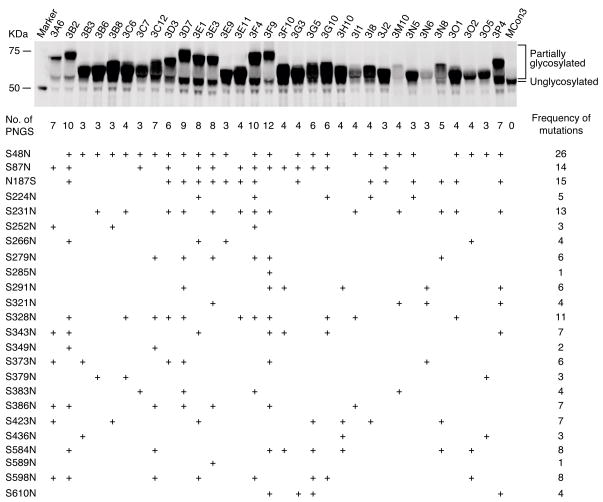
Figure 1.
Identification of clones containing introduced mutations by Western blot and sequencing analysis. The same amount (0.5 μg) of each plasmid DNA was transfected into 293T cells. Equal volume (12 μl) of culture supernatant was loaded into NuPAGE® Novex 4–12% Bis-Tris gels. Anti-HIV-1 Env mAb 13D5 and Alexa Fluor® 680 conjugated goat anti-mouse IgG were used to detect Env proteins. The image was acquired using the Odyssey Infrared Imaging System (Li Cor Bioscience, Lincoln, NE). The clones with added PNLGs showed increased molecular weight of the expressed Env Proteins. The mutation sites introduced in individual clones were confirmed by sequence analysis.

