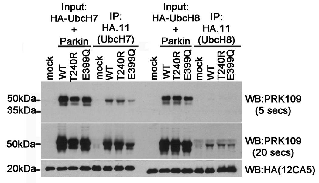
Figure 9.
The E399Q Parkin mutant shows reduced binding to UbcH7 and UbcH8. HEK293T cells were co-transfected with pRK5-HA-UbcH7 or pRK5-HA-UbcH8 and either pcDNA3.1 mock vector (“mock”) or with untagged full-length WT, T240R, or E399Q human parkin pcDNA3.1 constructs. Cells were harvested and soluble protein lysates were extracted (“Input”). The cell lysates were then immunoprecipitated using the anti-HA polyclonal antibody, HA.11 (“IP”). Input and IP fractions were resolved by SDS-PAGE and analyzed by immunoblot with the monoclonal antibodies PRK109 and anti-HA (clone 12CA5). The PRK109 blots are shown at 5 second and 20 second exposure times to highlight the differences in signal intensity reflected by the binding of parkin with UbcH7 versus that with UbcH8.

