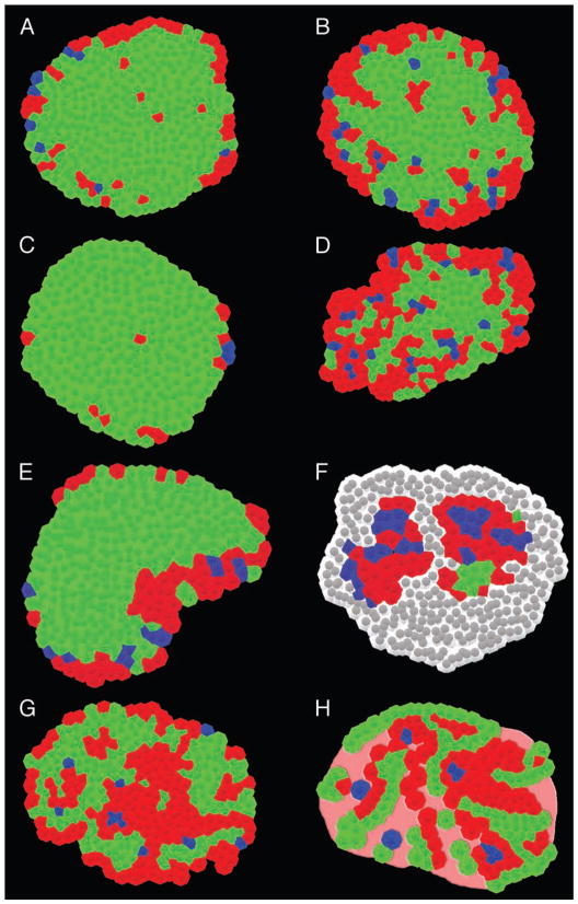
Figure 2.
Comparison of mouse and human islets under various pathophysiological conditions. Mouse and human islets under various pathophysiological conditions are shown; α-cells (red), β-cells (green), and δ-cells (blue) based on immunohistochemical images. (A) Wild-type mouse (6 mo). (B) Pregnant mouse (3 mo). (C) ob/ob mouse (15 wk). (D) db/db mouse (15 wk). (E) Neonatal mouse (P14). (F) NOD mouse (40 wk). Note that lymphocytes (shown in gray) infiltrate the islet replacing the majority of the endocrine cells.15 (G) Normal human (41 yr).16 (H) Type-2 diabetic human.40 Note that islets from patients with type 2 diabetes often contain acellular amyloid deposits as shown in pink.

