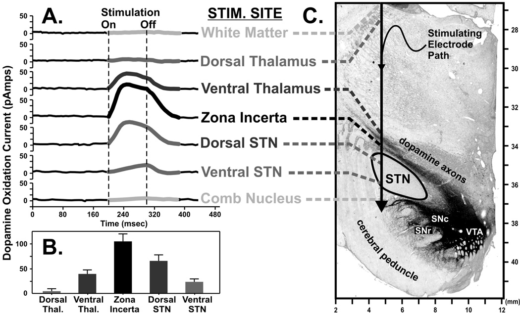
FIGURE 8.
Dopamine release measured in the dorsomedial tail of the caudate nucleus of the awake monkey (A) in response to electrical stimulation applied to various anatomical locations in and around the subthalamic nucleus (STN) as depicted in a representative coronal section of the monkey midbrain. Graph of the relationship of maximal increases in dopamine release with respect to the location of the stimulating electrode (B). Tyrosine hydroxylase-immunostaining highlights catecholaminergic axons originating in the dopamine-containing cell body regions of the substantia nigra pars compacta and reticulata (SNc and SNr, respectively) and ventral tegmental area (VTA) and can be seen coursing dorsally and through the STN on their way to the caudate (C).

