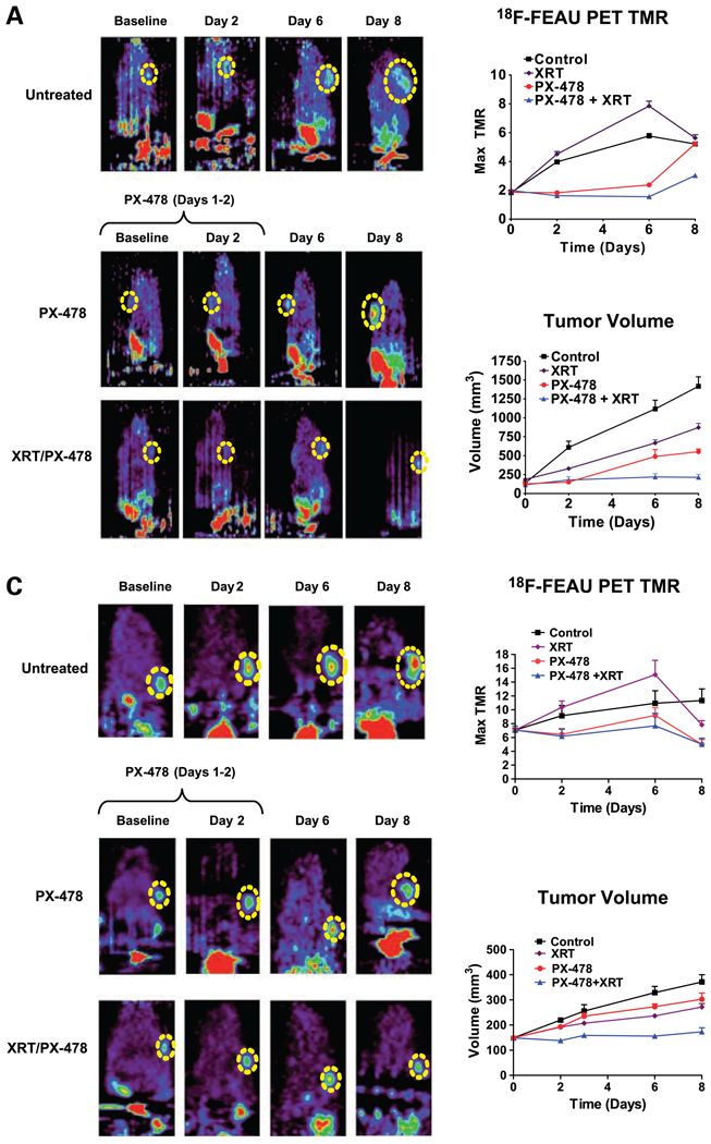
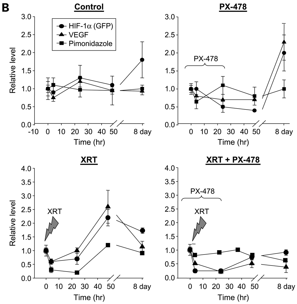
Figure 3.
PX-478 inhibits tumor HIF-1 signaling in vivo and markedly enhances growth delay in C6 and HN5 xenografted tumors following single-fraction radiation. A, 18F-FEAU PET imaging of C6 reporter xenografts (n = 8–10 in each study cohort) treated with 30 mg/kg PX-478 with or without 8 Gy single-fraction irradiation (XRT). Each series of images was obtained from the same animal at indicated time points. PX-478 was administered on days 1 and 2 of the time course, after baseline images had been obtained. Dashed yellow circle, location of each tumor on PET images. Animals are oriented with head at the top of each image; nonspecific bladder/kidney signal is seen below due to urinary excretion of 18F-FEAU. Points, mean values for maximum TMR of 18F-FEAU accumulation; bars, SE (top right). Points, mean values for tumor volumes across time; bars, SE (bottom right). B, C6 reporter xenograft tissue was harvested at indicated time points for immunohistochemical staining for HIF-1–dependent GFP expression (●), downstream VEGF expression (▲), and hypoxia-dependent retention of pimonidazole (■). Points, mean values (n = 4 tumors × 5 tissue sections) of expression levels normalized to baseline values; bars, SE. C, 18F-FEAU PET imaging of HN5 reporter xenografts (n = 6 per cohort) treated with 30 mg/kg PX-478 with or without 8 Gy single-fraction irradiation. Points, mean values for maximum TMR and tumor volumes, plotted as in A; bars, SE.


