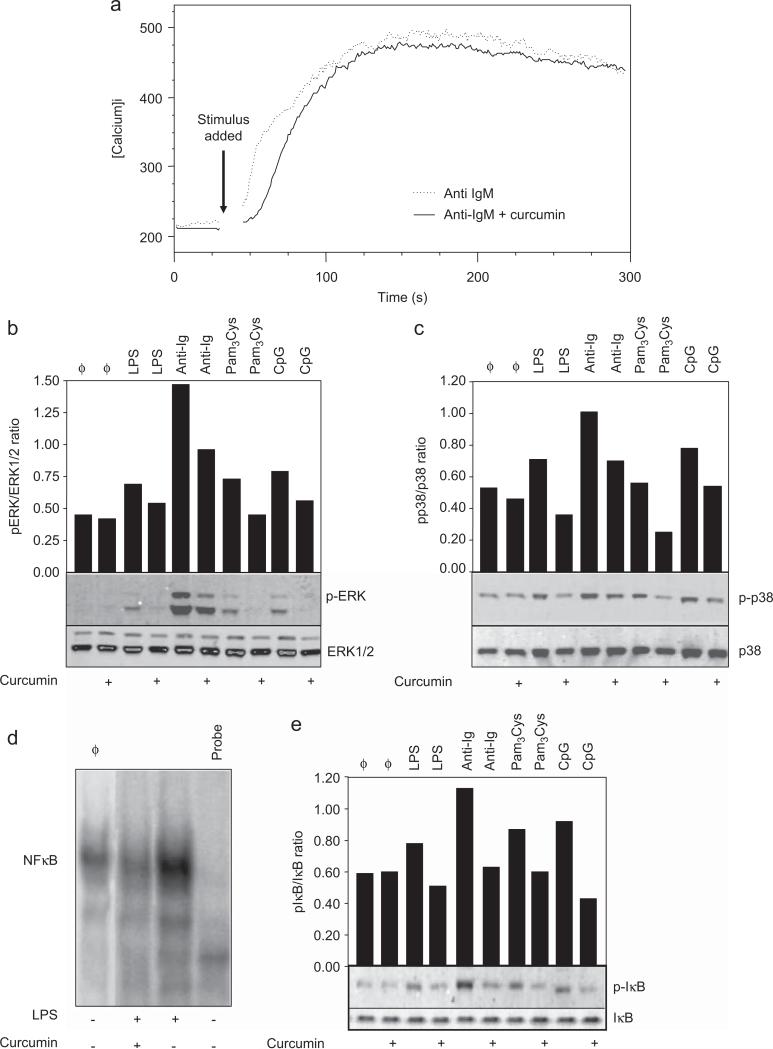
Fig. 3. Effect of curcumin in B cell signaling transduction.
(a) Intracelular calcium levels. Purified splenic murine B cells were loaded with Indo-AM and then stained with anti-B220-FITC antibody. Baseline calcium levels were determined by flow cytometry and the cells were then stimulated with anti-IgM (10 μg/ml) and treated with curcumin (10 μM). The mean calcium levels were determined by measurement of the fluorescence ratio of Indo-1-AM (390 nm/490 nm emission) and analyzed by the FlowJo software. Data are representative of three independent experiments. (b and c) MAPK phosphorylation. Purified B cells were stimulated with LPS (10 μg/ml), CpG oligodeoxynucleotides (1 μg/ml), Pam3Cys (1 μg/ml) or anti-IgM antibody (10 μg/ml). Some cultures were simultaneously treated with curcumin (10 μM). Cells were then lysed after either a 5 min (anti-IgM-treated cells) or a 30 min (TLR ligands-treated cells) treatment and whole-cell lysates were loaded onto SDS-PAGE gels. Blot were run and probed with the following antibodies: (b) anti-phospho ERK antibody; (c) anti-phospho-p38 antibody. The same blots were then stripped and reprobed with antibodies to nonphosphorylated proteins to determine absolute protein levels. Bar graphics shows the ratio of phosphorylated and total proteins. Data are representative of five independent experiments. (d) NFκB translocation to the nucleus. Purified B cells were treated with curcumin (10 μM) and stimulated with LPS (10 μg/ml) overnight. Nuclear extracts were prepared and tested for NFκB binding activity by EMSA using a double-stranded NF-κB consensus oligonucleotide. A double-stranded mutated oligonucleotide was used to examine the specificity of NFκB binding. The specific NF-κB bands are indicated. (e) IκB phosphorylation. B cells were stimulated with LPS (10 μg/ml), CpG (1 μg/ml), Pam3Cys (1 μg/ml) or anti-IgM antibody (10 μg/ml). Curcumin was added at 10 μM where indicated. Cells were then lysed after either 5 min (anti-IgM-treated cells) or 30 min (TLR ligands-treated cells) treatment and whole-cell lysates were loaded onto SDS-PAGE gels. Blot were run and probed with anti-phospho-IκB antibody. The same blots were then stripped and reprobed with anti-IκB antibody to determine absolute protein levels. Bar graphics shows the ratio of phosphorylated and total proteins. Data shown are representative of three independent experiments.

