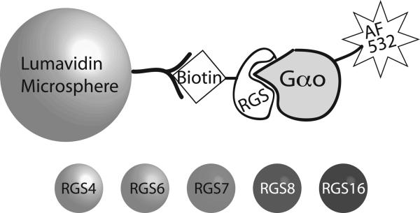
Figure 1.
Diagram of the components of FCPIA. Avidin-coated microspheres are incubated with biotinylated RGS protein to yield RGS-coated beads. 5 different bead regions are utilized, with each RGS being bound to a unique bead region. These bead regions are discriminated in the Luminex analyzer, and associated fluorescently labeled Gαo is detected. An inhibitor would result in the disruption of the RGS/Gαo interaction, and detected as a loss of bead-associated fluorescence.

