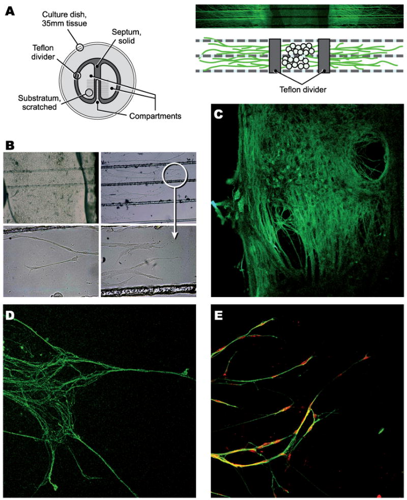
Figure 2.

Compartmented sensory neuronal cultures. A: schematic and ß-III tubulin immuno-stained cultures; B: phase contrast images of middle and side chambers (arrow head: growth cone); C-D: TuJ1 immuno-staining of neurons and distal axons; E: Schwann cells (red colored) ensheating axons in side chambers.
