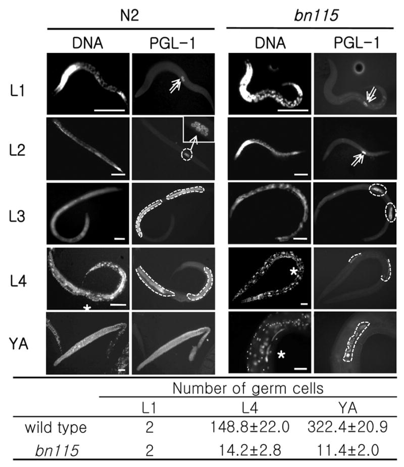
Fig. 2.

Loss of function of cdc-25.1 causes a germ cell proliferation defect. Postembryonic germline development was observed by immunostaining with anti-PGL-1, a germline-specific marker, along with Hoechst nuclear staining. Left: Wild-type N2, Right: cdc-25.1 (bn115) mutant. (A–D) At L1 stage, both N2 and bn115 worms have two primordial germ cells (PGCs), Z2 and Z3 (arrows). (E–H) At L2 stage, N2 contains around 10–20 germ cells (see magnified view in F), but bn115 still contains only 2 germ cells because of delayed cell divisions. (I–L and M–P) At L3 and L4 stages, germline mitotic proliferation continues in N2, producing hundreds of germ cells, which are circled with dotted lines, while the numbers of germ cells are much smaller in bn115 gonads. (Q–T) At the adult stage, N2 gonads display a progression of germline development from distal to proximal, including mitotic region, transition zone, pachytene region, oocytes in diakinesis, and sperm, while bn115 gonads contain only small numbers of undifferentiated germ cells. The N2 gonad shown (Q and R) was dissected out of the body. (U) Numbers of germ cells per gonad arm in cdc-25.1(bn115) hermaphrodites at different postembryonic stages. Numbers of PGL-1 positive germ nuclei per gonad arm in hermaphrodite worms of wild-type N2 and cdc-25.1 (bn115) mutant at each developmental stage are shown. Only for L1 stage, numbers are given per worm and not per gonad arm, as the two gonad arms in a hermaphrodite are not separated until the L3 stage. For each value, 10 hermaphrodite gonad arms were examined by fluorescence microscopy. L1, first larval stage; L4, fourth larval stage; YA, young adult stage, in which worms had already finished L4-to-adult molting but had not started laying eggs yet. Numbers are presented as mean ± SD. In each panel, anterior is left and dorsal is up. Asterisks indicate the position of vulva. Scale bars, 50 μm.
