
Figure 2.
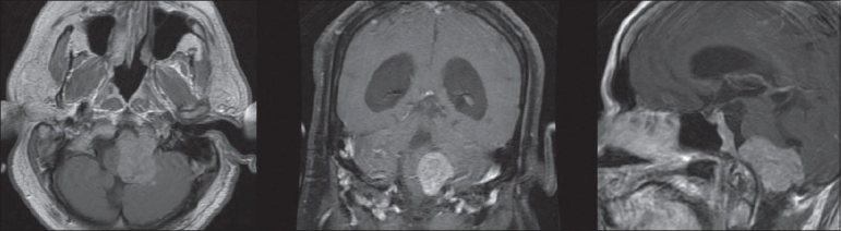
Preoperative MRI of the brain T1-weighted postcontrast infusion sequences in axial, coronal, and sagittal planes demonstrating an extra-axial, eccentrically shaped mass with contrast enhancement in the left aspect of the medullary cistern compressing and displacing the brainstem and cerebellum to the right