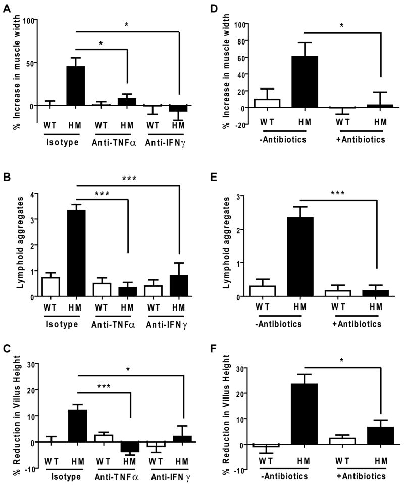
Figure 6. TNFα and IFNγ inhibition or antibiotics treatment ameliorates DSS-induced disease in virus-infected Atg16L1 mutant mice.
(A) Isotype control or blocking antibodies against TNFα and IFNγ were administered to WT and Atg16L1HM mice infected with MNV CR6 for 7 days followed by additional 7 days of DSS treatment at which point intestines where harvested. The muscularis propria thickness in the region adjacent to the ano-rectal junction was quantified and normalized to the average of WT mice receiving isotype control antibodies (n ≥ 6 mice/condition, *p < 0.05, mean +/−SEM).
(B) Number of lymphoid aggregates in the region adjacent to the ano-rectal junction from mice in (A) (*p < 0.001, mean +/−SEM).
(C) Quantification of average villus height from mice in (A) normalized to the average of WT mice receiving isotype control antibodies (an average of 263 total villi from at least 5 mice/condition was measured, *p < 0.05, ***p < 0.001, mean +/−SEM).
(D) WT and Atg16L1HM mice were orally inoculated with MNV CR6 for 7 days and then given 2.5% DSS and broad spectrum antibiotics concurrently for another 7 days at which point intestines where harvested. The muscularis propria thickness was normalized to the average of uninfected WT mice treated with DSS from Figure 4D (n = 6 mice/condition, *p < 0.05, mean +/−SEM).
(E) Number of lymphoid aggregates from mice in (D) (***p < 0.001, mean +/−SEM).
(F) Quantification of average villus height from mice in (C) normalized to the average of uninfected WT mice treated with DSS from Figure 5B (an average of 320 total villi from at least 5 mice/condition was measured, *p < 0.05, mean +/−SEM).
See also Figure S5.

