
Figure 4.
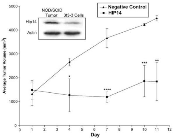
Intratumoral injections of HIP3 siRNA. Tumors were established in female NOD/SCID mice by subcutaneous injections in the right flank with 1 × 10⁶ 3t3-3 cells. At 28 days postinjection, the tumors were measured and the animals were split into two groups with roughly equal tumor volumes. HIP14 or negative control siRNAs (30 μM) were injected intratumorally on days 1, 4, 7, and 10, at which time tumor volumes were measured. A statistical comparison of the two groups at days 4, 7, 10, and 11 showed significant differences for all four points, with the following P-values; *P>0.0273, ****P>0.0007, ***P>0.0034, **P>0.0045 as determined by unpaired t-tests. Inset is a Western blot analysis of tumor tissue and 3t3-3 cells