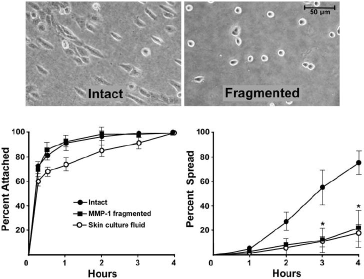
Fig. 2.

Attachment and spreading of human epidermal keratinocytes on intact and fragmented collagen lattices. Left panel: attachment. A similar rate of attachment is observed on intact and fragmented collagen. Right panel: spreading. This is significantly reduced on fragmented collagen. Values are means and standard deviations based on triplicate samples in a single experiment. Statistical significance was determined using the Student t-test, comparing each treatment group to its control. ‘*’ indicates statistical difference from control at P < 0.05 level. The experiment was repeated five times with similar results. Inset: appearance of keratinocytes after 2 h on intact and fragmented collagen lattices
