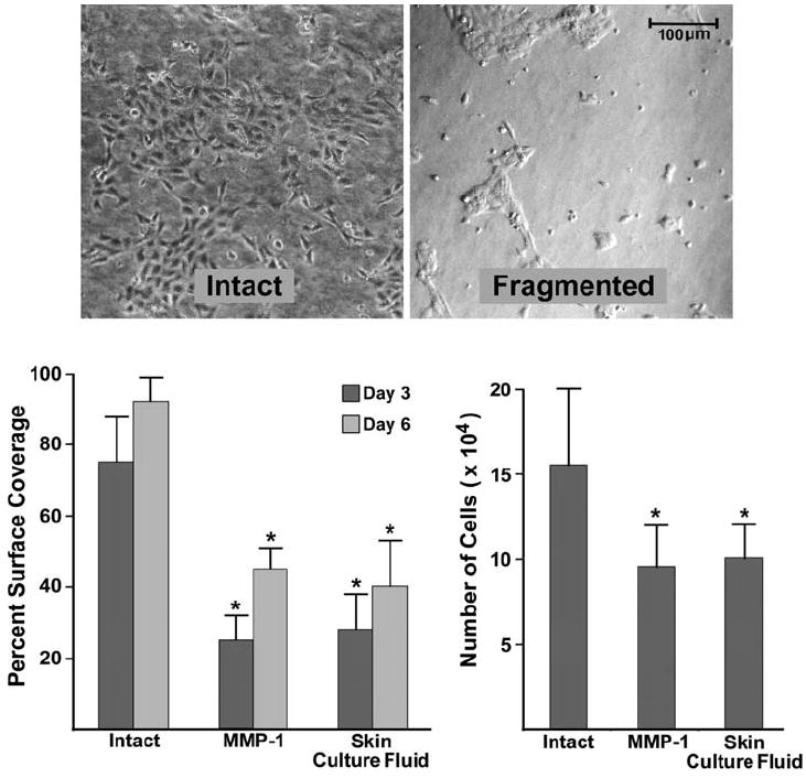
Fig. 3.

Keratinocyte coverage of the surface of intact and fragmented collagen lattices. Left panel: percentage of the collagen surface covered with cells on Days 3 and 6. Values shown are means and standard errors based on seven different experiments. Right panel: proliferation. Cells were harvested and counted on Day 3. Values shown are means and standard errors based on four separate experiments. Statistical significance was determined using the Student t-test. ‘*’ indicates statistical difference from control at P < 0.05 level. Inset: appearance of keratinocytes after 3 days on intact and fragmented collagen
