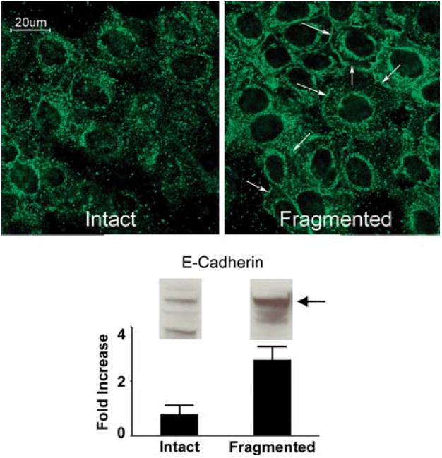
Fig. 4.

E-cadherin expression in keratinocytes on intact and fragmented collagen. Upper panels: confocal immunofluorescence microscopy of human epidermal keratinocytes after 48 h on a three-dimensional polymer of intact collagen or fragmented collagen. E-cadherin expression is more diffuse in the cells on the intact collagen substrate. E-cadherin is localized to the cell surface to a greater extent on damaged collagen. Expression is especially prominent at sites of cell-to-cell contact (arrows). Lower panel: western blot for E-cadherin. Increased E-cadherin was detected in cells on damaged collagen. Values shown in the bar graph represent digitized bands in western blots from two separate experiments and represent means and ranges. Confocal microscopic analysis was done with three separate preparations
