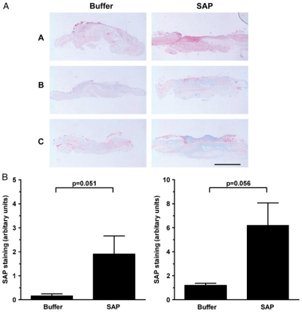
Figure 8.
(A) Detection of serum amyloid P (SAP) in SAP-treated wounds 1–3. Photographs of wound sections stained with anti-SAP antibody. Note increased SAP expression (pink stain) in SAP-treated wounds compared with corresponding control wounds. 1: Intradermally treated wounds harvested at post-wounding day 4, 2: Intradermally treated wounds harvested at post-wounding day 7 and 3: Intraperitoneally treated wounds harvested at post-wounding day 7. Bar = 2mm. (B) Significantly increased % SAP staining area was noted in both the intradermally treated wounds (left) and intraperitoneally treated wounds (right) compared with corresponding control wounds.

