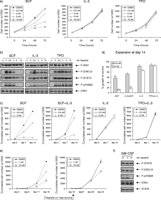
Fig. 3.

Dasatinib selectively impairs SCF-induced and mutant KIT-driven signal transduction and proliferation. a Mo7e cell line was cultured in RPMI with 5% FCS, supplemented with 20 ng/ml SCF, IL-3, or TPO, in the absence or presence of increasing concentrations of dasatinib. Average expansion of three independent experiments is shown. b Mo7e cells were deprived of cytokine in RPMI with 0.5% FCS overnight, followed by the pretreatment with dasatinib for 2 h. The cells were harvested for lysates after being left unstimulated (lane U) or stimulated with 20 ng/ml SCF, IL-3, or TPO for 15 min. Cells (3 × 10⁵) were loaded for Western blot, analyzing with antibodies as indicated. c Freshly isolated CB CD34+ cells (3 × 10⁴) were cultured in IMDM with 10% FCS, supplemented with SCF (20 ng/ml), IL-3 (20 ng/ml), or in combination (SCF+IL-3), as well as TPO (20 ng/ml) and IL-3 (5 ng/ml) (TPO+IL-3). Demi-depopulation was performed at days 7 and 14 for counting. A representative experiment out of two independent experiments is shown. d Percentages of growth after dasatinib treatment as compared to control group (% growth of control) are shown as mean value from two independent experiments. e SKNO-1 cells (105) were plated in 12-well plates precoated with MS5 stromal cells. Cells were grown in RPMI supplemented 10% FCS, in the presence or absence of GM-CSF (10 ng/ml). Dasatinib was added as indicated concentrations. Cultures were demi-depopulated as indicated time points for analysis. Weekly cumulative cell counts represented cells in suspension. The leukemic cells in suspension were harvested from the coculture at day 16 to initiate the second cocultures on new MS5 stroma. f SKNO-1 cells were cultured in RPMI supplemented 10% FCS, in the presence of GM-CSF (10 ng/ml), with dasatinib treatment for 4 h. Cell lysates were prepared and subjected to Western blotting using antibodies as indicated
