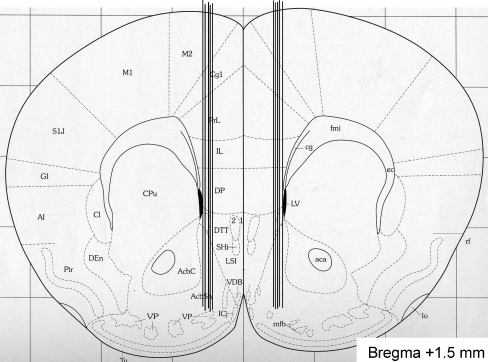
Fig. 3.
Verification of probe placement. A coronal mouse brain section showing ten representative probe placements (vertical lines) in the NAcc of mice used in the present study (Franklin and Paxinos 1996). Ten representative placements are illustrated, but all other placements were within the NAcc shell. The probe is not shown to scale, and the outer diameter of the probe was 310 μm. Placements outside this area were not included in the statistical analysis. The number given in the brain section indicates millimeters anterior (+) from bregma

