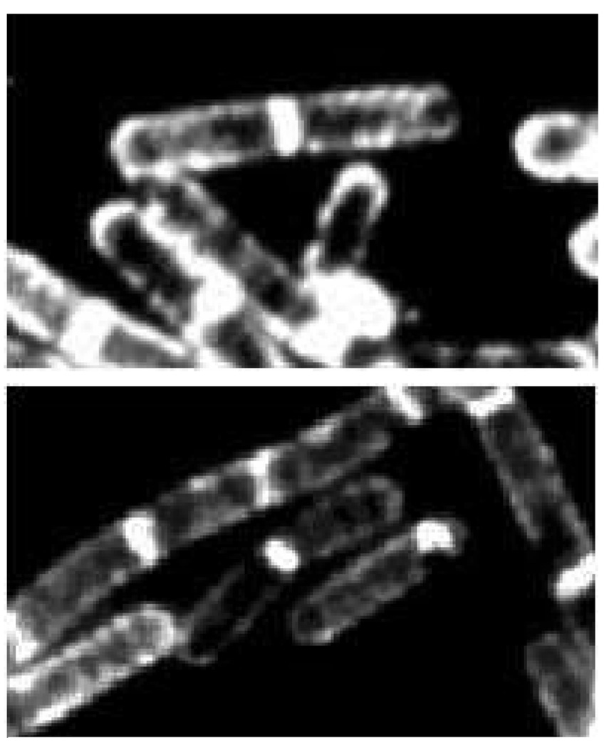
Figure 1.
Visualization of the external peptidoglycan surface of Gram-positive bacteria by the use of fluorescently-labeled peptidoglycan-binding antibiotics. These fluorescent microscopy images, reproduced from the study by Tiyanont et al. Proc. Natl Acad. Sci. U. S. A. 2006, 103, 11033–11038 © 2006 National Academy of Sciences, show rod-shaped B. subtilis (the bacterium is approximately 2 µm in length) bacteria stained with fluorescein-labeled ramoplanin. The peptidoglycan is intensely stained at the newly forming division septum, and to a lesser extent on the sidewalls and the old pole. These locations coincide with the sub-cellular locations of Lipid II, the key biosynthetic precursor of the peptidoglycan (see Scheme 1). The sidewall staining is suggestive of a helical pattern for peptidoglycan growth during sidewall elongation.

