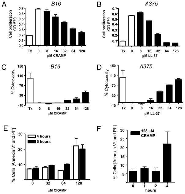
FIGURE 3.
Cathelicidin peptides inhibit melanoma cell proliferation and induce LDH release and melanoma cell lysis in vitro. Murine B16 or human A375 melanoma cells were coincubated for 24 h with different concentrations of recombinant LL-37 or mCRAMP peptides, respectively. A and B, Cell proliferation was evaluated by a modified MTT assay and recorded as absorbance at 570 nm. C and D, Cell death was quantified by measuring LDH release from damaged cells and is plotted as percent cell death relative to Triton X-100 (Tx)–treated cells used as positive controls. Data are means + SD from triplicate measurements. One representative experiment out of three is shown. E and F, Annexin V and PI staining were performed on B16 cells that were incubated with recombinant mCRAMP peptides. E, Concentrations of mCRAMP that induced LDH release from B16 cells also showed double-positive Annexin V+ and PI+ staining. F, Double-positive Annexin V+ and PI+ cells were seen within 4 h of incubation with 128 μM CRAMP. Data shown are percentage of cells that were Annexin V+ and PI+ double positive.

