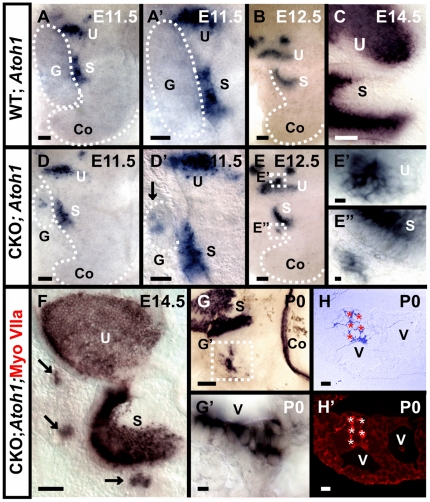
Figure 2. Persistent Atoh1 expression in remaining ganglia relates to transformation of ganglionic cells into hair cells in Neurod1 mutant.
In situ hybridization of Atoh1 shows a faint and transient expression at E11.5 in some vestibular ganglion cells in wild-type mice (A, A’). This expression is more profound and continues in the ganglia of Neurod1 CKO mice (D, D’). In later stages, Atoh1 in situ signal appears in a cluster of cells in CKO mutants near the utricle and saccule (E–E’, F, G–G’). Some of the Atoh1 positive cells are aligned along the vesicular lumen similar to the Myo VIIa positive cells shown in Fig. 1 (G). To investigate co-localization, we labeled Atoh1 in situ reacted ears with anti-Myo VIIa antibody, embedded in plastic and sectioned. The sections reveal co-localization of Myo VIIa with Atoh1 in these cells (H,H’) thereby providing strong evidence that these cells are hair cells. S, saccule; U, utricle; G, ganglia; V, intraganglionic vesicle. Bar indicates 100 µm except E, E’ and 10 µm in E, E’.

