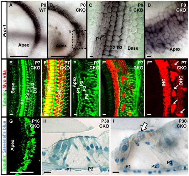
Figure 6. Neurod1 regulates organization of supporting cells as well as hair cells.
A supporting cell marker, Prox1, shows uniform expression in two rows of pillar and three rows of Deiter’s cells in wild-type mice (A) and in the base of Neurod1 CKO mice (B,C). In contrast, the apex of Neurod1 CKO mice shows multiple disorganized rows of supporting cells with no clear distinction between Pillar and Deiter’s cells (D). At P7, combined tubulin and Myo VIIa immunocytochemistry shows normal organization of supporting cells in between hair cells in the base of the mutant cochlea (E, E’). This orientation is disrupted in the apex where Myo VIIa positive hair cells are surrounded by supporting cells with thick processes filled with tubulin. This is reminiscent of Pillar cells and not of the thinner phalangeal processes of Deiter’s cells (F, F”, G). Detailed histology in thin plastic sections of P30 mutant cochlea show not only the persistence of multiple rows of inner and outer hair cells in the apex (H) but also the formation of multiple rows of Pillar cells (P1, P2, P3) and inner hair cells in places of outer hair cells (arrow in I). These data suggest that Neurod1 is an important transcription factor that mediates the coordinated type specific hair cell and supporting cell development in the apical half of the organ of Corti which is necessary for the coordinated development of supporting cells. P1, P2, inner and outer Pillar cells; D1, D2, D3, 1st, 2nd, 3rd row of Deiter’s cells; IHC, inner hair cells; OHC, outer hair cells. Bar indicates 100 µm except C, D, H, I and 10 µm in C, D, H, I.

