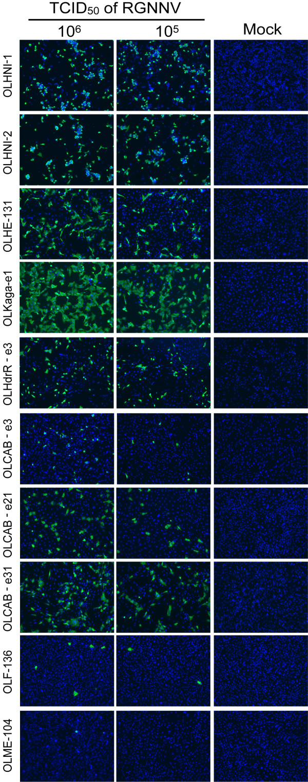
Figure 1.
Infectivity of RGNNV in various medaka cell lines. Each cell line (1.0-1.5 × 10⁵ cells) was inoculated with 105 or 106 TCID50 of RGNNV and incubated at 30°C. Viral coat protein in infected cells was detected by indirect immunofluorescence assay at 1 dpi. Cell nucleus was stained with 4', 6-diamino-2-phenylindole (DAPI). Data represents the merged image of Alexa488-fluorencence and DAPI staining.

