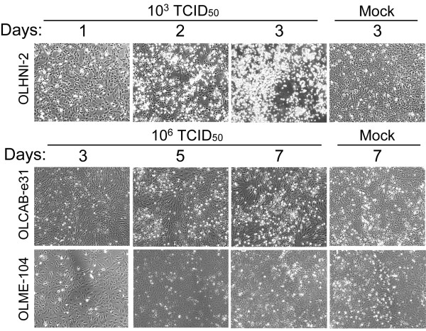
Figure 2.
CPE development in RGNNV-infected medaka cells. The cells (1.0-1.5 × 10⁵) were inoculated with RGNNV of the indicated titers and cultured at 30°C. Cell morphology of the RGNNV-inoculated or mock-inoculated cells was observed at 1-3 dpi for OLHNI-2 cells and at 3-7 dpi for OLCAB-e31 and OLME-104 cells.

