Figure 3.
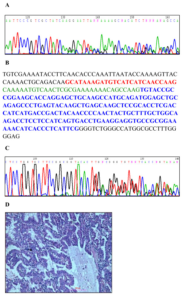
Concurrent heterozygous ALK fusion and EGFR deletion in a single adenocarcinoma. (A) Chromatograph indicating a heterozygous deletion (2235-2249 del 15) in exon 19 of EGFR in a patient with EML4-ALK. (B) Sequence of EML4-ALK fusion variant 3b. Red sequence indicates exon 6 of EML4. Green sequence indicates an insertion of 33 bp from intron 6 of EML4 found in variant 3b. Blue sequence indicates exon 20 of ALK. (C) Chromatograph for EML4-ALK fusion variant 3b indicating the presence of a heterozygous fusion of ALK with exon 6 of EML4 with an additional 33 bp insertion. (D) Histological staining of the adenocarcinoma from the patient with both an EGFR mutation and EML4-ALK fusion. Scale bar = 100 μm.

