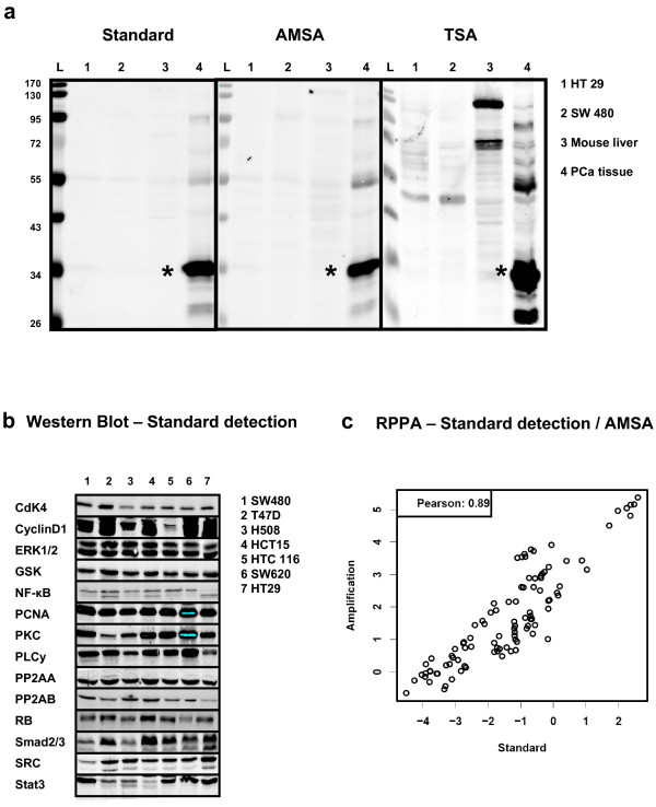
Figure 3.
Comparing standard NIR detection and AMSA. (a) Western blot specificity of TSA and AMSA. Detection of PSA (star-symbol) using standard NIR procedure (scan intensity 5), antibody-mediated signal amplification (scan intensity 2.5) and TSA (scan intensity 2.5). Each lane corresponds to loading 5 μg total protein from prostate cancer tissue or PSA-free negative controls: Colon cancer cell lines HT29 (1), SW480 (2), mouse liver lysate (3), and prostate cancer tissue (4). (b) Western blot analysis of 14 proteins in seven human cancer cell lines with standard NIR detection. (c) Samples were analyzed by RPPA to compare standard NIR detection and AMSA using the same set of antibodies as shown for the Western blot. The correlation analyses were based on log signal intensities.

