
Figure 2.
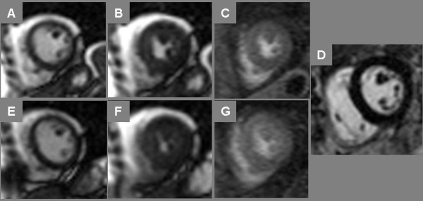
Normal Treadmill Stress CMR. End-diastolic (A, E) and end-systolic (B, F) frames of cine imaging at rest (top row) and immediately post-stress (bottom row) plus stress myocardial perfusion imaging (C, G) are shown in a 52 year-old postmenopausal female referred for stress SPECT to evaluate dyspnea; both stress modalities were negative for ischemia. In addition, late post-gadolinium enhancement (LGE) CMR imaging (D) showed no myocardial enhancement.