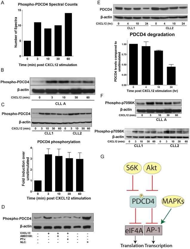
Figure 5. CXCL12 Induces Phosphorylation of PDCD4 at Ser457.
A) Bar graph depicting the spectral counts of PDCD4 phosphopeptides observed in the LC-MS/MS analysis at time points of CXCL12 stimulation. B) Western blot of PDCD4 phosphorylation over time course of 0 to 60 min CXCL12 stimulation (30 nM) from CLL A patient cells. β-actin served as a loading control. C) Top panel: Representative western blot of PDCD4 phosphorylation in CLL cells from 2 different CLL patients not used in LC-MS/MS analysis over 30 nM CXCL12 stimulation time course. β-actin served as a loading control. Bottom panel: Densitometry analysis of PDCD4 phosphorylation levels CXCL12 stimulation (30 nM) time points relative to unstimulated controls and averaged from 10 separate CLL patient cells. Error bars represent standard error of the mean (SEM). D) Western blot of PDCD4 phosphorylation in unstimulated/untreated CLL cells or 3 min CXCL12 stimulations (30 nM) in the presence (+) or absence (−) of preincubation (1 h) with AMD3100 (40 µM) or Pertussis toxin (PTx) (200 ng/ml). NLC lysate represents CLL cells cultured in presence of NLCs with no further stimulation or treatment. CLL cells were removed from the adherent NLCs and lysed. β-actin served as a loading control. E) Top panel: Representative western blot detecting total levels of PDCD4 in CLL cells following 0, 4, 10 or 24 h of 30 nM CXCL12 stimulation. β-actin served as a loading control. Bottom panel: Bar graph quantifying total PDCD4 levels over 24 h time course of 30 nM CXCL12 stimulation compared to 0 h unstimulated controls and normalized to β-actin levels by densitometry analysis of western blots. Data represented are mean +/− SD of 3 separate CLL patients' cells. F) Western blot stripped and reprobed from Figure 4B for p70S6K phosphorylation (Thr389) over time course of 30 nM CXCL12 stimulation from CLL A patient cells (Top panel) and 2 other representative CLL patients' cells (bottom panel). β-actin served as a loading control. G) Diagram of PDCD4 signaling showing known upstream regulators as well as downstream targets. Akt and p70S6K are known to phosphorylate PDCD4, thereby inhibiting its function in repressing eIF4A translational activity and AP-1 transcription.

