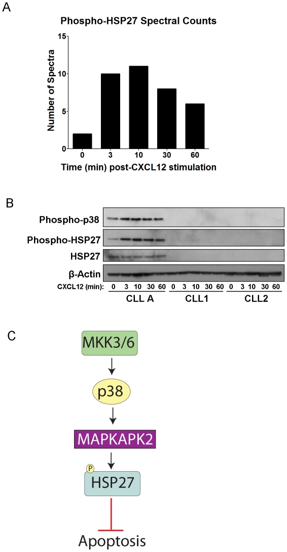
Figure 6. Phosphorylation of HSP27 in Subset of CLL Patients.
A) Bar graph depicting the spectral counts of HSP27 phosphopeptides (Ser82) observed in the LC-MS/MS analysis after CXCL12 stimulation. B) Western blot detecting phosphorylation of HSP27 and the upstream p38-MAPK, and total HSP27 over time course of 0 to 60 min CXCL12 stimulation (30 nM) from CLL A patient cells and 2 other representative CLL patients' cells. β-actin was run as a loading control. C) Signaling diagram of HSP27, which can protect from apoptosis, and its upstream regulation by p38-MAPK and MAPKAPK2.

