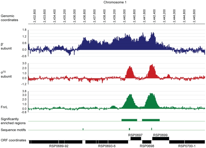
Figure 5. Identification of FnrL binding sites in the R. sphaeroides genome by ChIP–chip assays.
A representative region of the R. sphaeroides genome showing profiles resulting from the enrichment of DNA fragments by immuno-precipitation of the β′ subunit (blue) or σ70 (red) subunit of RNA polymerase or FnrL (green) is plotted along the indicated genomic coordinates. The data plot the log2 of the ratio of the immunoprecipitated sample to the control sample as a function of probe location along the genome (coordinates are indicated in base pairs). DNA regions significantly enriched (p-value ≤0.01) by FnrL immuno-precipitation (green boxes), positions of sequences matching the FnrL consensus binding site (green tick mark) and the coordinates of annotated genes (black boxes). The data were plotted using SignalMap 1.9 (NimbleGen Systems).

