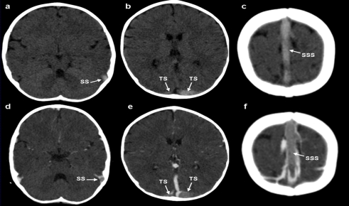
Figure 1.
Unenhanced axial computed tomography (CT) (a-c) shows the hyperdense appearance of the left sigmoid sinus (SS), left and right transverse sinus (TS), and the superior sagittal sinus (SSS), representing acute thrombus (“cord sign”). Enhanced axial CT images (d-f) obtained at the same levels showing the filling defects within the cerebral veins corresponding to sinus vein thrombosis.

