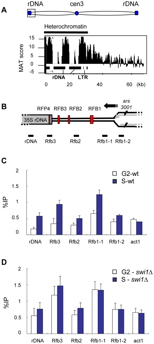
Figure 5. γH2A is highly enriched in the rDNA repeats during S phase.
(A) Detailed ChIP-on-chip distribution of γH2A in the rDNA on the left arm of chromosome 3. Black rectangles (below graph) represent 35S rDNA gene repeats. (B) Diagram of one rDNA repeat (not to scale) shows the location of the four replication fork barriers (red vertical bars) relative to the 35S rDNA genes, the direction of replication (black arrow) from the ars3001 replication origin, and qPCR primer locations, below graph. (C, D) γH2A ChIP at the rDNA was performed in wild type and swi1Δ strains synchronized by cdc25-22 block and analyzed by qPCR with the indicated primers.

