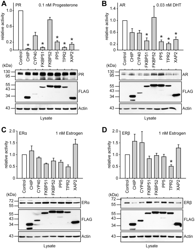
Figure 4. PR, AR, ERα and ERβ activities in the presence of different TPR proteins.
SK-N-MC cells were transfected with the MMTV-Luc (for PR and AR assays), or the ERE-Luc reporter plasmid (for ERα and ERβ assays), the Gaussia-KDEL control plasmid, a plasmid expressing the HA-tagged steroid hormone receptor as indicated and the plasmid expressing a FLAG-tagged TPR-protein. After transfection, cells were cultivated for 24 h in the presence of hormone as indicated. Relative receptor activity represents firefly data normalized to Gaussia activities and presented as relative stimulation to control + S.E.M. of at least four independent experiments performed in duplicate. Control cells were transfected with cloning plasmid replacing the TPR protein expression plasmid in the transfection mixture. Lower panels of A–D display immunoblots of cell extracts, probed with anti-HA antibody visualizing steroid receptor expression, the same membrane probed with FLAG antibody demonstrating expression of the TPR proteins and with actin antibody as loading control. * denotes p-values ≤0.001.

