
Figure 4.
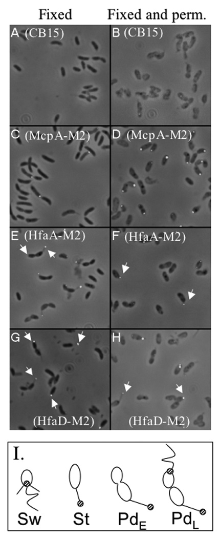
Localization of HfaA-M2 and HfaD-M2 using immunofluorescence microscopy. Panels A, C, E, and G fixed cells. Panels B, D, F, and H fixed and permeabilized cells. Panels A and B, CB15; Panels C and D, NA1000 mcpA::pRCM22; Panels E and F, CB15 hfaA::pJM23hfaA; Panels G and H, CB15 hfaD::pJM23hfaD. White arrows in panels E thru H indicate polar localization for HfaA and HfaD in swarmer, PD and stalked cells. (I) Cartoon of C. crescentus indicating the localization of HfaA and HfaD (striped circle) in swarmer (Sw), stalked (St), early PD (PdE) and late-PD (PdL) cells.