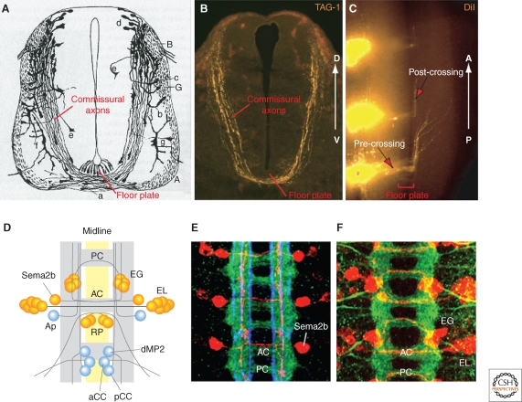
Figure 1.
Commissural axons at the midline in vertebrates and Drosophila. (A) Drawing by Cajal showing commissural axons by Golgi staining of E4 chick embryonic spinal cord. Note the growth cones at the tip of axons. (B) Commissural axons revealed by immunostaining with a marker, TAG-1, in an E11.5 mouse spinal cord transverse section (similar stage as E4 chick embryo). (C) Midline crossing and anterior turning of commissural axons revealed by DiI tracing in E11.5 mouse open-book spinal cord preparation. (D) Schematic of the ventral nerve cord of a late stage Drosophila embryo, anterior up. Yellow indicates midline cells, gray indicates axon tracts (adapted from Keleman et al. 2002). AC, anterior commissure; PC, posterior commissure. Examples of identified neurons and their projections are shown, with commissural neurons in orange and ipsilateral neurons in blue. Sema2b, an intersegmental commissural neuron; EG and EL, intrasegmental commissural neurons; RP, commissural motorneurons; dMP2, ipsilateral intersegmental neuron with posterior projections; pCC, ipsilateral intersegmental neuron with anterior projection; aCC, ipsilateral motorneuron. (E) Confocal image of the nerve cord, with all axons stained in green, specific longitudinal pathways in blue (anti-FasII), and Sema2b neurons in red (adapted from Rajagopalan et al. 2000b). (F) Confocal image of the nerve cord with all axons stained in green and EG and EL neurons in red (adapted from Brankatschk and Dickson 2006).

