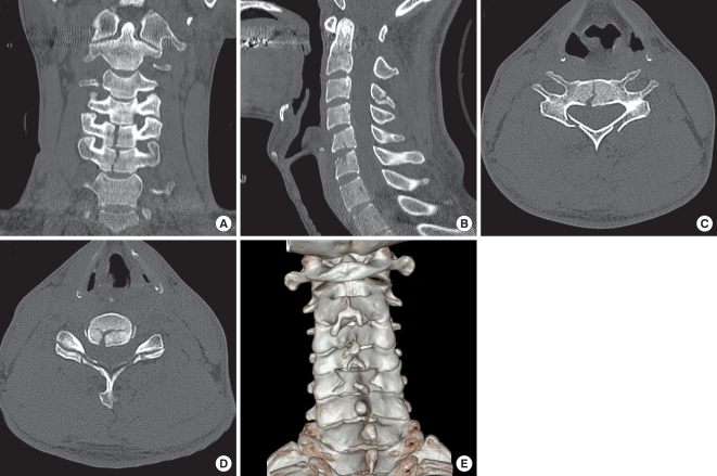
Fig. 2.
Cervical spine computed tomographic (CT) scans with 3-dimensional (3-D) reconstruction. (A) C5 and C6 vertebral bodies show vertical fracture centrums in a coronally reconstructed image. (B) Only subtle C5 retrolisthesis on C6 is noted on a sagittally reconstructed image. A C3 spinous process fracture is also identified. (C) The axial section of the C5 vertebra at the pedicle level shows a depressed laminar fracture. (D) The fracture lines of the C5 lamina are through the lamina-facet junction. The C5/6 facet joints are intact. (E) 3-D reconstructed image shows a C5 lamina depressed fracture through the lamina-facet junction.

