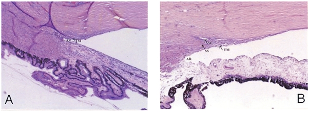
Fig. 3.
(A) Histopathologic view of an angle with delayed-onset aphakic glaucoma. There is no identifiable Schlemm's canal (SC), the scleral spur (SS) is indistinct, and the trabecular meshwork (TM) is compact. The angle recess (AR) is poorly developed (H&E, magnification, ×12). (B) An age-matched normal filtration angle of a 15-year-old female who received enucleation due to retinoblastoma (H&E, magnification, ×12).

